- 55 例原发性尿道腺癌分为新发与前列腺放疗后两类;放疗后多见于近距离粒子治疗后多年。
- 组织学谱含 NOS、肠型、黏液性、透明细胞、腺样囊性,并可肉瘤样变;部分放疗后病例 H3K27 缺失。
- 侵袭性强,但新发与放疗后及各亚型间 DSS 未见显著差异。
摘要
本文报告 55 例原发性尿道腺癌的临床病理与影像分析。确诊年龄 42–88 岁(平均 66 岁);男 26、女 29(1.0∶1.1)。分为两组:新发尿道腺癌(n=45)与前列腺腺癌放疗后尿道腺癌(n=10,放疗后腺癌)。放疗后肿瘤发生于放疗后 8–25 年(平均 15.6 年);8 例曾行近距离放疗。新发组 45 例中女 29 例(女∶男 1.8∶1.0);17 例女性肿瘤发生于尿道憩室。
组织学上表现为腺癌 NOS,或透明细胞、黏液性、肠型及腺样囊性形态。2 例新发与 3 例放疗后进展为肉瘤样癌。
全队列平均疾病特异性生存 100.35 月。Kaplan–Meier 曲线显示新发与放疗后组疾病特异性生存模式相似。
引言
原发性尿道腺癌极为罕见,略偏女性。先天性憩室与获得性狭窄是促成女性优势的易感因素。因罕见,文献多为个案或小样本系列,其中透明细胞腺癌报道最多。大样本综合报告极少。原发性尿道腺癌的临床与形态谱仍未完全界定,生物学行为亦了解不足。故本回顾性研究分析 55 例原发性尿道腺癌的临床病理特征,包括 10 例在前列腺腺癌放疗后继发肿瘤的患者。
材料与方法
组织样本
检索得克萨斯大学 MD Anderson 癌症中心病理档案中 1984 年 1 月至 2025 年 4 月全部原发性尿道腺癌。复核病理报告、HE 切片及相关免疫组化。原发性尿道腺癌定义为起源于尿道、呈纯腺样分化的肿瘤;排除膀胱腺癌或伴腺样分化的尿路上皮癌继发累及尿道者。由 3 位泌尿生殖专科病理医生(M.Z.、P.T.、B.C.)复阅。评估大体外观、大小、原位成分、组织学特征、邻近器官/结构累及、脉管侵犯、淋巴结转移及远处转移,并与随访、治疗及影像等临床资料对照。
免疫组织化学染色
于 UT MD Anderson 进行下列免疫组化:CK7(Dako,1:100)、p63(Santa Cruz,1:1000)、CK20(Dako,1:40)、血栓调节蛋白(Dako,1:40)、CK5/6(Dako,1:100)、PIN 双染试剂(Biocare,1:40)、CDX2(BioGenex,1:900)、GATA-3(Leica,1:200)、SATB2(Cell Marque,即用)、K27MH3.3(Sigma-Aldrich,1:500)、NKX3.1(Cell Marque,1:100)、PAX8(Cell Marque,即用)。
按厂家说明行 HPV 原位杂交(Enzo)及 RNAscope HPV 检测(Advanced Cell Diagnostics)。FFPE 组织切 5±1 μm;脱蜡脱水后,于相邻三张切片分别加 HPV16/18、泛素 C 及 dapB 探针,扩增后 DAB 显色、复染封片。
临床资料
汇总年龄、性别、既往肿瘤史及临床/膀胱镜所见;随访与结局取自病历。经机构伦理批准后,由泌尿外科专家(C.P.)复核临床表现与治疗。分析疾病特异性生存(DSS),并绘制新发组与放疗后组 Kaplan–Meier 曲线比较。
影像学研究
35 例有影像资料(女 18、男 17)。MRI 28 例(男 16、女 12),CT 7 例(女 6、男 1)。MRI 在本院(n=13)或外院(n=15)完成;均可获得 T2 与 T1,24 例有增强序列。
结果
临床表现与病史
共 55 例——女 29、男 26(比例 1.1∶1.0),诊断时平均年龄 66 岁(42–88 岁)。临床、病理与影像资料见补充表 1。临床表现包括尿道狭窄、尿失禁、排尿梗阻、血尿、阴道瘙痒/分泌物或会阴痛。按病史分为两组:新发尿道腺癌(45 例)与前列腺腺癌放疗后尿道腺癌(10 例,称放疗后腺癌);放疗后判定遵循 Cahan 标准。新发组 45 例中女 29 例(女∶男 1.8∶1.0);其中 17 例女性肿瘤发生于尿道憩室。
放疗后尿道腺癌发生于放疗后 8–25 年(平均 15.6 年)。8 例近距离放疗,1 例外照射,1 例放疗类型不详。
大体所见
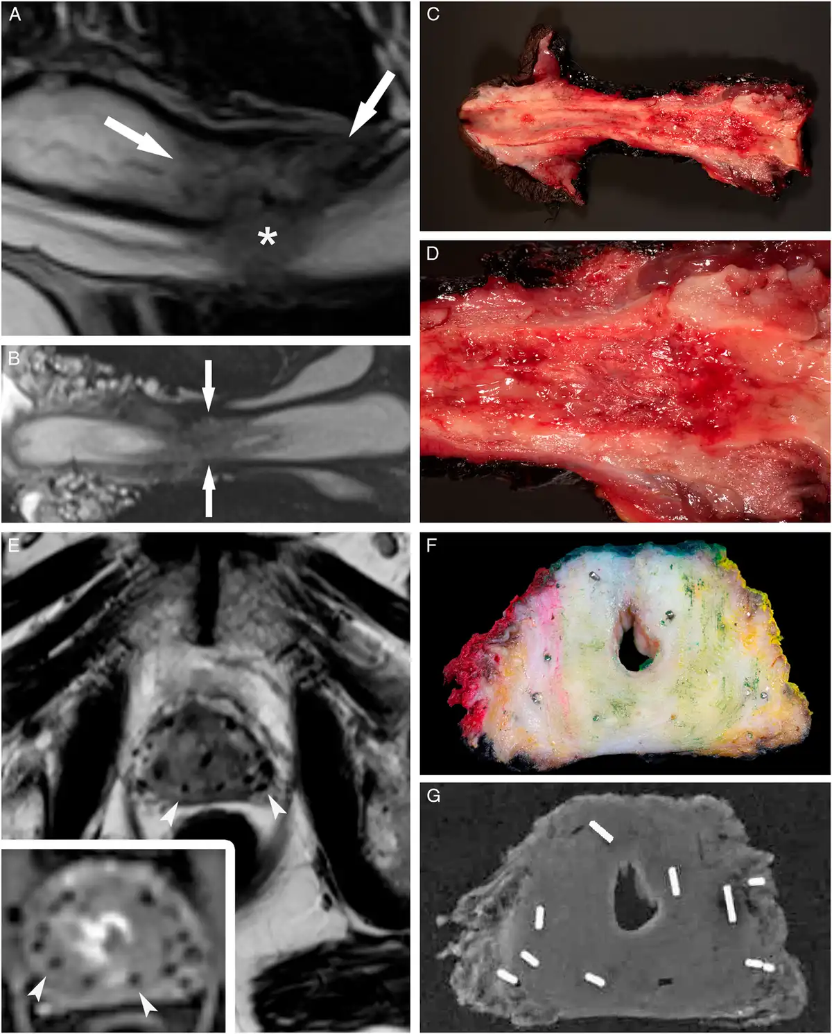
20 例有大体资料(新发 18、放疗后 2),并与影像对照(图 1A–G)。平均瘤径 3.2 cm(0.5–6.2 cm)。肿瘤多围绕尿道、质脆、边界不清(图 1A–G)。切面黄至灰,可黏液样,表面常粗糙。常浸润尿道周围软组织及前列腺、膀胱颈、阴道或海绵体。
图 1.
镜下所见
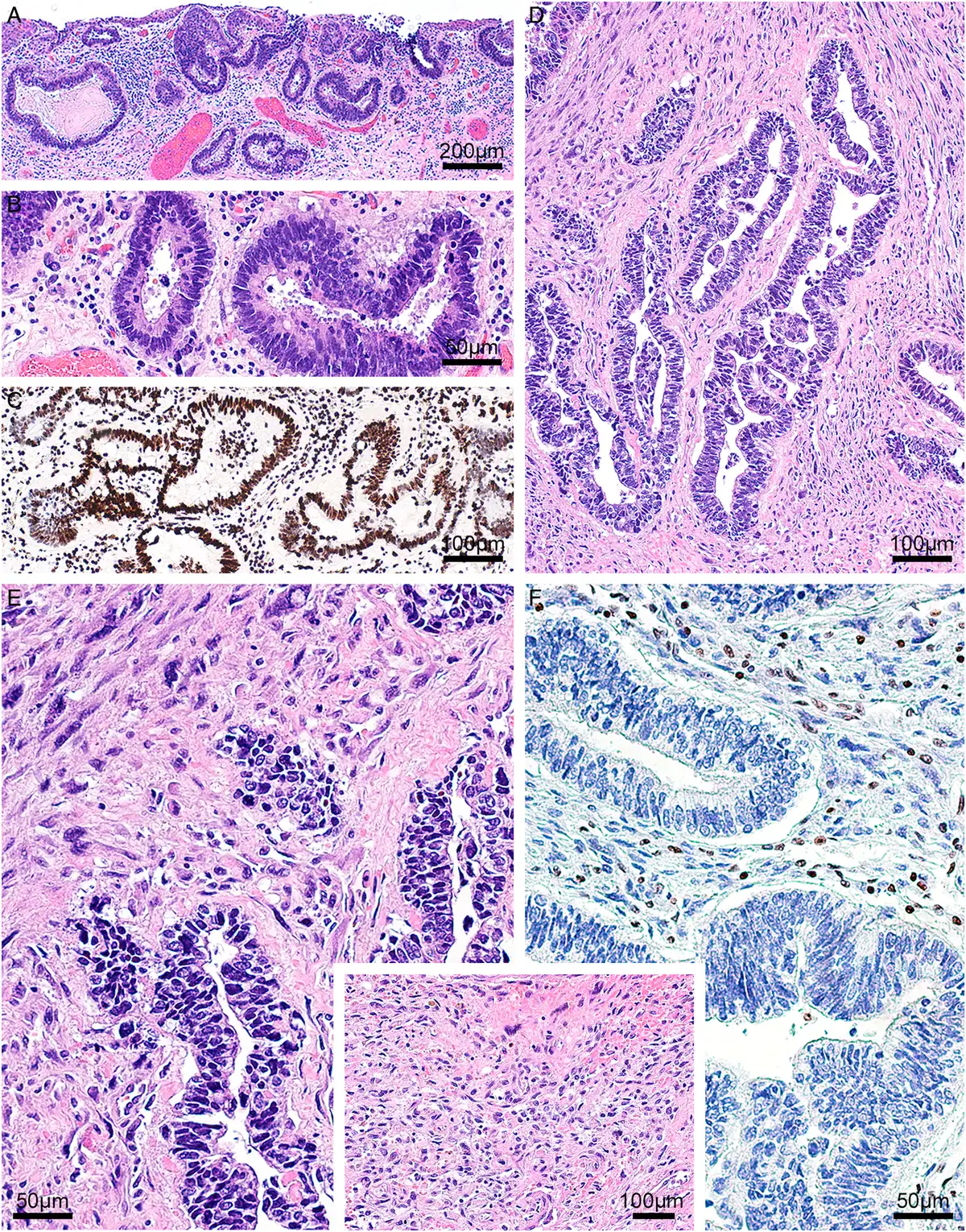
组织学上分多种亚型。新发组:非特殊类型腺癌(NOS;n=13)(图 2A,B)、肠型(n=11)(图 2C,D)、黏液性(n=8)(图 3A–C)、透明细胞(n=11)(图 3D,E)及腺样囊性(n=2)。2 例黏液性腺癌有显著印戒细胞成分;另 1 例同时含肠型腺癌与大细胞神经内分泌癌(图 4A–D)。
图 2.
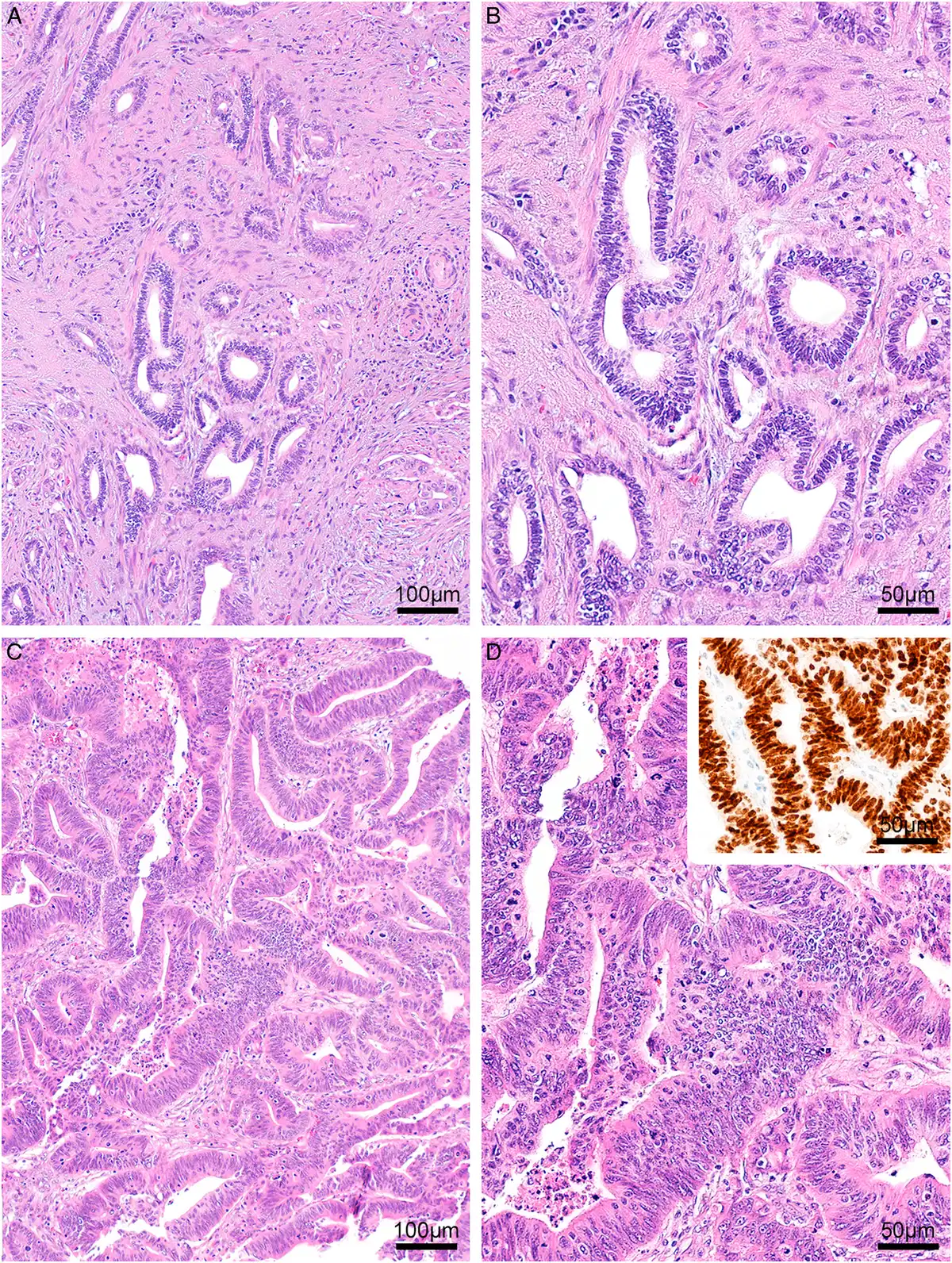
图 3.
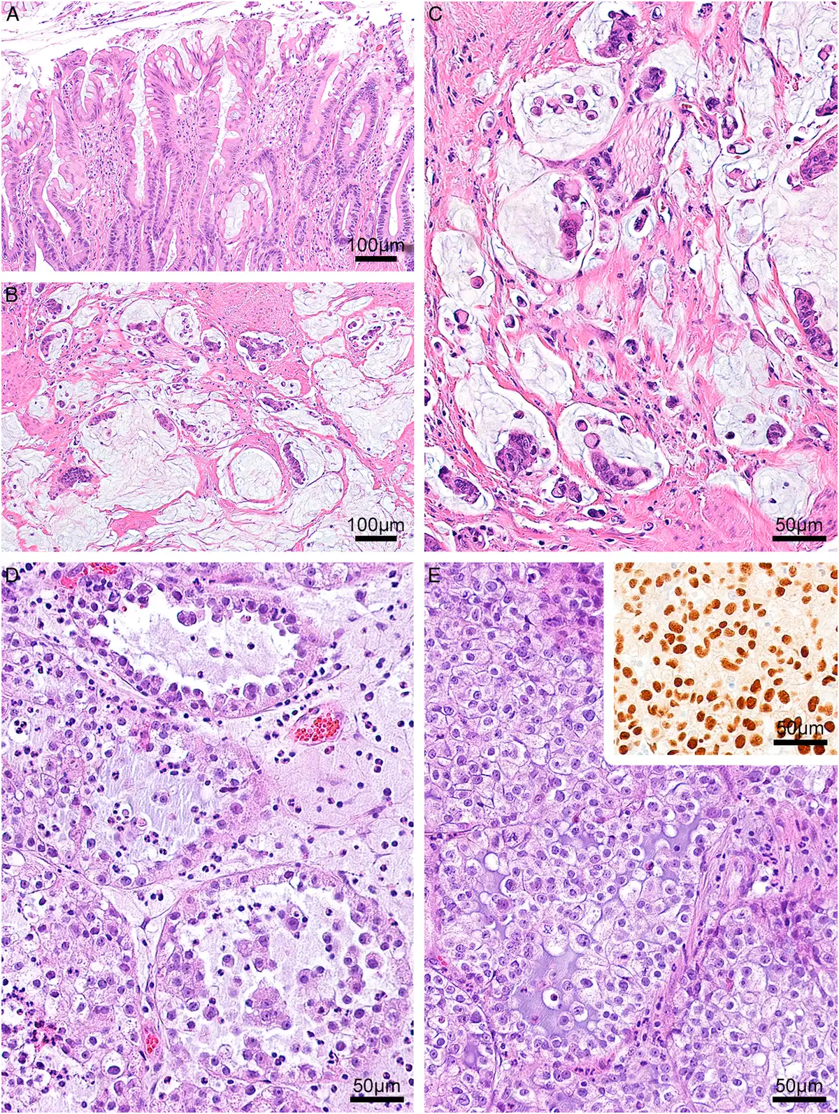
图 4.
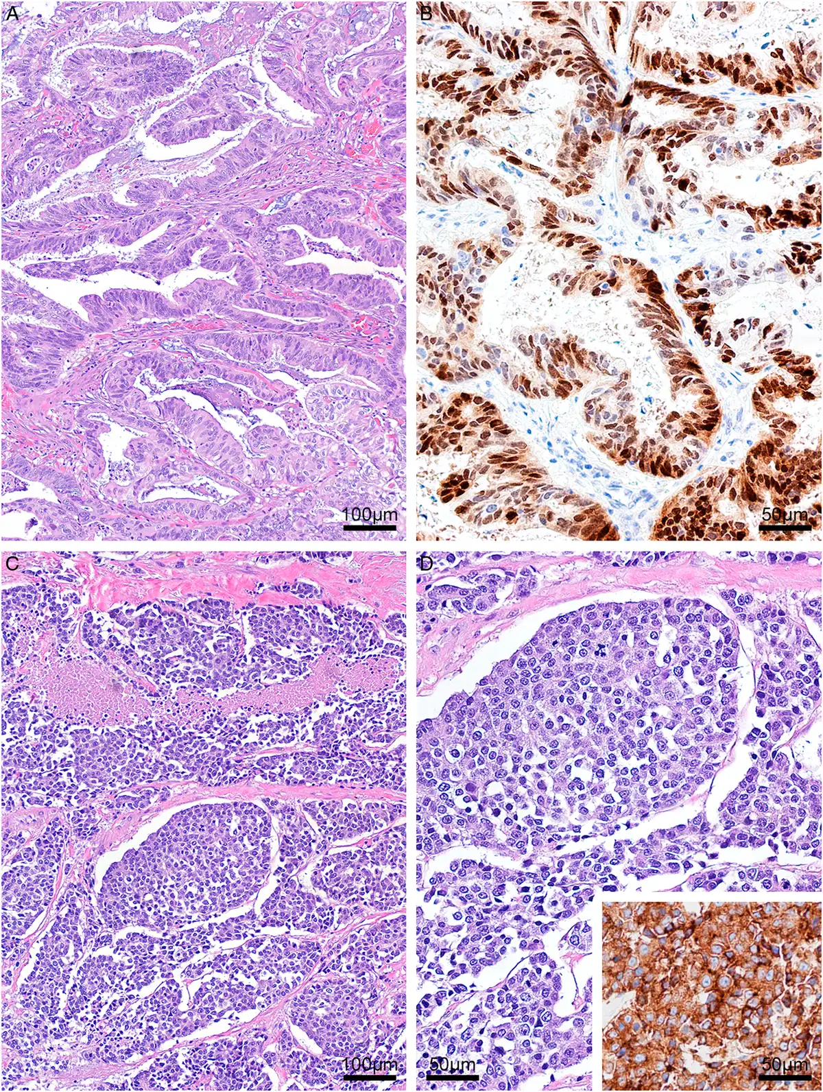
新发组透明细胞腺癌 PAX8 阳性(图 3E 插图)。非透明细胞腺癌免疫表型明显不同:多数 CK7(n=8)、SATB2(n=6)、CK20(n=6)阳性。所测病例均呈强弥漫核 CDX2 阳性(n=8,图 2D 插图)。CK5/6、p63 阴性(n=8);GATA-3 阴性(n=8);NKX3.1 阴性(n=20)。3 例 HPV 原位杂交(黏液性 2、肠型 1)高危 HPV 均阴性。混合肠型与大细胞神经内分泌癌中,神经内分泌成分 synaptophysin 强弥漫阳性(图 4D 插图);该例 PSA、PSAP、NKX3.1 两成分均阴性以排除前列腺来源。2 例腺癌 NOS 进展为肉瘤样癌,呈高级别未分化梭形细胞及多形性肉瘤特征。
放疗后腺癌包括腺癌 NOS(n=4)(图 5A–D)、肠型(n=2)、肠型+黏液性(n=1)及黏液性(n=3)。其中 3 例进展为肉瘤样癌(图 6A–F)。全部放疗后腺癌上皮样成分核 CDX2 阳性。
图 5.
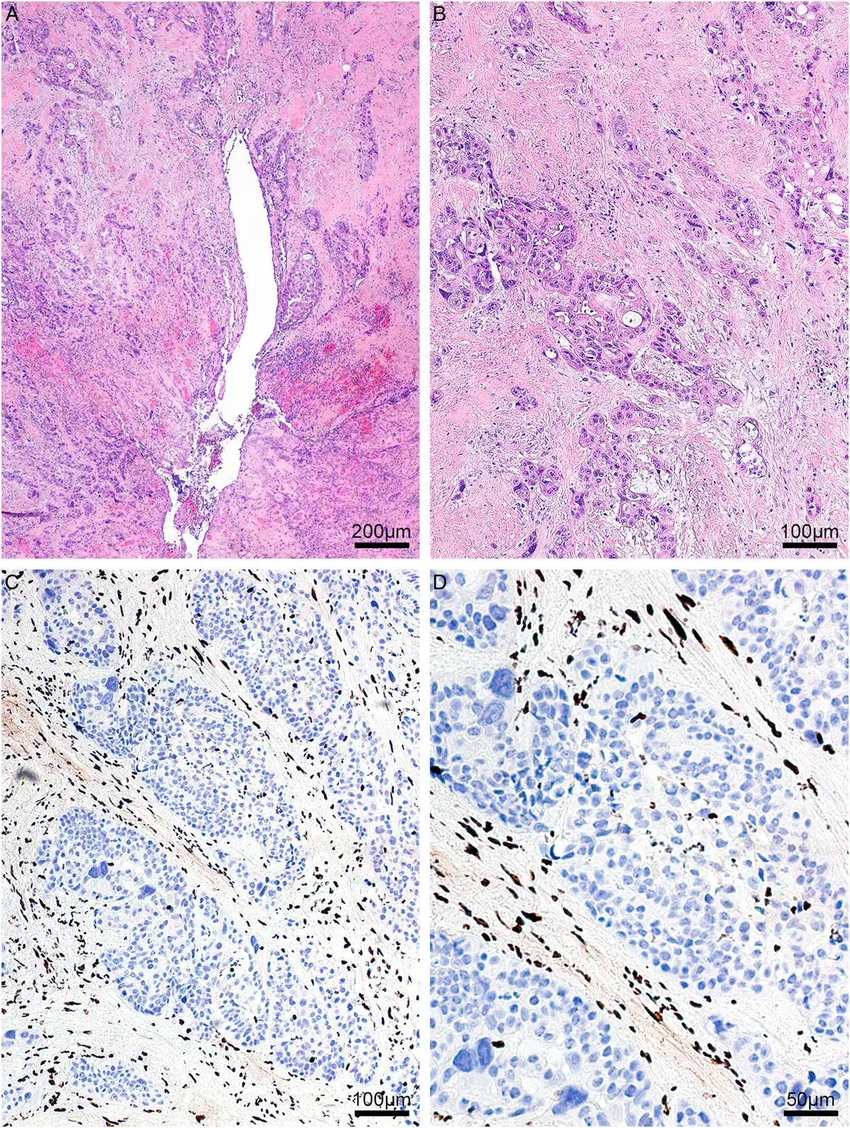
图 6.
2 例放疗后腺癌 NOS 见组蛋白 ME3H3(H3K27)缺失,其中 1 例进展为肉瘤样癌——放疗后肉瘤常见现象(图 5C–D、6F)。肉瘤样放疗后腺癌中,肉瘤样与浸润性腺癌成分均缺失甲基化组蛋白 ME3H3(H3K27);同例邻近覆盖尿道黏膜内原位腺癌仍保留 ME3H3(H3K27)染色。
另见 2 例新发腺癌在邻近覆盖尿道黏膜有原位腺癌(图 7A–F);均发生于腺性化生背景上,含高度异型腺样结构及多层腺样表面增生。
图 7.
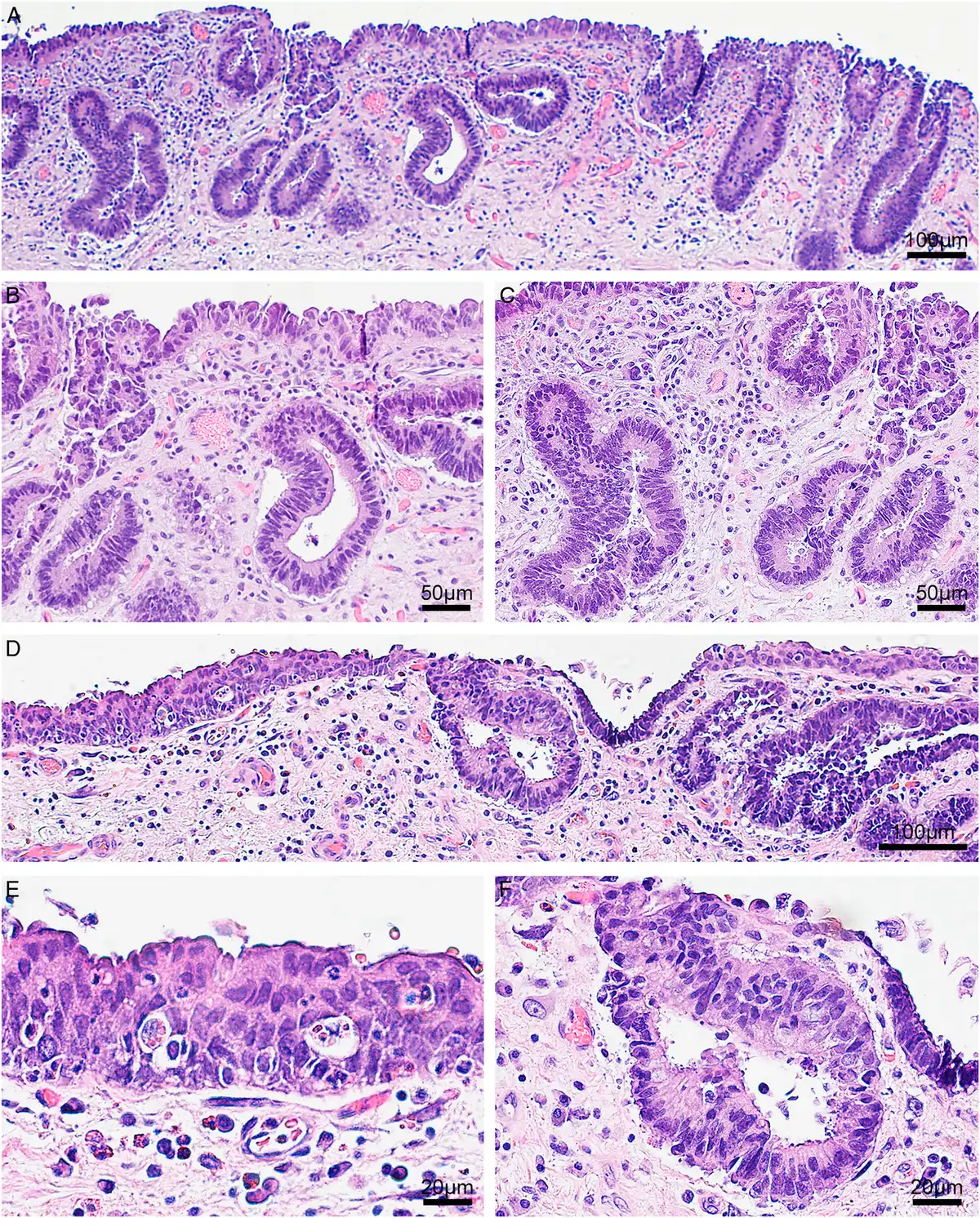
影像学所见
35 例有影像。新发组 28 例亚型分布:肠型 7、黏液性 4、透明细胞 7、NOS 8、腺样囊性 2。
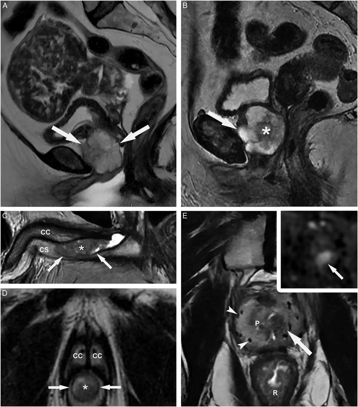
新发组女性中半数以上肿瘤发生于尿道憩室(图 8A,B 及补充图)。男性 9 例起自前列腺部尿道,亦可累及球部/膜部尿道(n=8)及阴茎尿道(n=6)。
各亚型影像特征显著重叠。肠型腺癌 T2 常低信号,增强早期明显强化并常侵及邻近结构。腺癌 NOS 亦常 T2 低信号、强化明显,倾向巨块或浸润性形态(图 8C,D)。黏液性肿瘤信号多变——男性常 T2 低、女性常高——强化不均、弥漫浸润(图 8A)。透明细胞癌以 T2 高信号、T1 等至高信号为特征,边界清、膨胀性、分叶伴分隔,多起自憩室(图 8A,B)。
透明细胞癌形态最具特征:分叶、分隔、边界清楚;几乎均见于女性,本组有 1 例男性。肠型与 NOS 较其他亚型更具浸润性,常累及前列腺、尿道海绵体、膀胱、阴道。黏液性肿瘤常不均质、弥漫浸润,偶有钙化。罕见腺样囊性癌影像非特异、低信号,见于男性膜部尿道。
放疗后腺癌 10 例中 7 例有影像:肠型 2、黏液性 1、肠型+黏液性 1、NOS 3(其中 2 例 NOS 进展为肉瘤样癌)。这 7 例均呈 T2 低信号,侵及前列腺间质并扭曲前列腺解剖、推移近距离放疗粒子(图 8E 及插图)。
图 8.
治疗与随访
12 例有转移信息(新发 11、放疗后 1)。新发组:5 例仅盆腔淋巴结转移;4 例淋巴结+器官转移(腹股沟/盆腔淋巴结合并肝、肺、阴道壁或皮肤);另 2 例仅远处转移(肝或骨)。放疗后组 1 例盆腔淋巴结转移。
32 例有治疗信息。新发组 24 例:术前化疗 6、放化疗 6、单纯化疗 4、单纯手术 3、放疗后手术 2、单纯放疗 1、放化疗后手术 1、放化疗+手术后免疫治疗 1。放疗后组 8 例:单纯手术 2、化疗后手术 2、单纯化疗 1、手术后化疗 1、同步放化疗 1、新辅助化疗免疫 1。
51 例有随访。新发组 41 例(平均 111 月,中位 65 月):无病生存 14、带病生存 9、死于本病 10、死于他因 1、存活但疾病状态不详 1、死因不详 6。放疗后组 10 例均有随访(平均 43 月,中位 32 月):无病生存 2、带病生存 5、死于本病 2、存活状态不详 1。
全队列平均疾病特异性生存 100.35 月,中位 55.53 月。新发组平均/中位 DSS 115.72/74.14 月;放疗后组较短(40.29/29.43 月),但差异无统计学意义。可能混杂因素包括新发组 7 例死于不明/他因,以及两组 DSS 死亡时间模式相似(Kaplan–Meier,补充图 S3)。亚型间生存差异亦无统计学意义(补充表 S2;两两比较 P 0.3–0.9)。随访资料不支持新发与放疗后尿道腺癌、以及不同病理亚组在临床侵袭性上存在显著差异。
讨论
原发性尿道腺癌极罕见,男女均可,略偏女性(本组女∶男 1.1∶1.0)。文献多为个案或小系列。本组临床、病理与影像显示两类:新发,或前列腺腺癌放疗后。新发组女性优势明显(1.8∶1.0),58% 女性肿瘤与尿道憩室相关。放疗后尿道腺癌仅见于男性,多为近距离粒子放疗后遗症,常于放疗后多年发生(平均约 15 年)。据我们所知,这是放疗后尿道癌的首个综合性报告。
镜下分为透明细胞与非透明细胞。本组透明细胞腺癌几乎均见于女性,部分伴既有憩室。非透明细胞包括肠型、NOS、黏液性,偶见腺样囊性。部分进展为肉瘤样癌。放疗后背景下肉瘤样癌可缺失组蛋白 H3K27,此为恶性外周神经鞘瘤标志,亦见于放疗后肉瘤。
鉴别诊断包括男性前列腺癌、邻近器官肿瘤累及甚至转移。需结合临床、影像与病理排除胃肠道来源的孤立性肠型/黏液性转移。继发累及的膀胱尿路上皮癌伴腺样分化可凭临床及腺样结构保留尿路上皮表型(GATA3 阳性)排除。罕见纯膀胱肠型/黏液性腺癌需结合膀胱镜与影像。结直肠与妇科继发灶靠病史与影像排除。男性黏液性/导管型前列腺癌靠前列腺特异性标志阴性排除。尚需与近期报道的、多数高危 HPV16/18 阳性的原发性尿道癌鉴别。覆盖黏膜原位癌最支持尿道起源,但仅见于少数病例。
影像虽不能特异对应组织学亚型,但有助于确认肿瘤以尿道为中心,支持尿道起源。
泌尿道(含尿道)透明细胞腺癌类似女性生殖道常见者;组织发生不清,曾提出苗勒管与中肾来源。分子上常有 ARID1A、TP53 突变,亦有 PIK3CA、PTEN 报道。相反,肠型/黏液性非透明细胞腺癌常有 KRAS、EGFR 突变。
随访显示尿道腺癌侵袭性强:全队列疾病特异性死亡平均/中位时间 31.28/26.53 月。新发与放疗后总体行为无显著差异(P=0.94);各病理亚型间亦无显著。但须承认治疗异质性与可分析例数有限为混杂因素。
补充材料
补充表与补充图见出版社 Supplemental Digital Content 链接(PAS/C411–C415)。正文未另附结构化数据表;图 1–8 已嵌入。
Abstract
Herein, we report the results of clinicopathologic and radiographic analyses of 55 patients with primary adenocarcinoma of the urethra. Their ages at diagnosis ranged from 42 to 88 years (mean: 66 y). Twenty-six patients were male and 29 were female (ratio: 1.0:1.1). We noted 2 distinct groups of cases: de novo urethral adenocarcinoma (n=45) and urethral adenocarcinoma in patients after radiation therapy for prostatic adenocarcinoma (n=10), defined as postradiation adenocarcinoma. Postradiation tumors developed 8 to 25 years (mean: 15.6 y) after radiation therapy. Eight patients underwent brachytherapy. Among the 45 patients with de novo adenocarcinoma, 29 were female, for a female-to-male ratio of 1.8 to 1.0. In 17 female patients with de novo urethral adenocarcinoma, tumors developed in a urethral diverticulum. Histologically, urethral adenocarcinomas exhibited adenocarcinoma not otherwise specified or clear cell, mucinous, enteric, and adenoid cystic carcinoma morphology. In 2 cases of de novo adenocarcinoma and 3 cases of postradiation adenocarcinoma, the tumors progressed to sarcomatoid carcinoma. The mean disease-specific survival time for the entire cohort was 100.35 months. As reflected by the Kaplan-Meier curves for these patients, the patterns of disease-specific survival were similar in the de novo and postradiation groups.
Introduction
Primary adenocarcinoma of the urethra is a very rare tumor, with a slight predilection for female individuals. 1–4 Congenital diverticula and acquired strictures are predisposing factors contributing to the female predominance of urethral adenocarcinomas. 5 Because of the rarity of this tumor, most reports of it in the literature are case reports or small series, with clear cell adenocarcinoma being the most common reported subtype. 6–15 Comprehensive reports of larger series are exceedingly rare. 1,2 The clinical and morphologic spectrum of primary urethral adenocarcinoma remains incompletely defined, and its behavior is poorly understood. Therefore, in this retrospective study, we analyzed the clinicopathologic features of 55 cases of primary adenocarcinoma of the urethra, including 10 patients in whom tumors developed as a sequela of radiation therapy for prostatic adenocarcinoma.
Materials and Methods
Tissue Samples
The pathology archives of The University of Texas MD Anderson Cancer Center were searched for all primary urethral adenocarcinoma cases from January 1984 to April 2025. Pathology reports, hematoxylin- and eosin-stained slides, and pertinent immunohistochemical studies were reviewed. Primary urethral adenocarcinomas were defined as tumors arising in the urethra with pure glandular differentiation. Cases with secondary involvement of the urethra by urinary bladder adenocarcinoma or urothelial carcinoma with glandular differentiation were excluded. The cases were reviewed by 3 dedicated genitourinary pathologists (M.Z., P.T., and B.C.). Tumors were evaluated for gross appearance, size, in situ components, histologic features, adjacent organ/structure involvement, lymphovascular invasion, lymph node metastasis, and distant metastasis. The pathologic data were correlated with clinical data, including follow-up, treatment, and imaging data.
Immunohistochemical Staining
Immunohistochemical studies to detect the expression of the following were performed at UT MD Anderson: CK7 (Dako, Carpinteria, CA; diluted 1:100), p63 (Santa Cruz Biotechnology, Dallas, TX; diluted 1:1000), CK20 (Dako; diluted 1:40), thrombomodulin (Dako; diluted 1:40), CK5/6 (Dako; diluted 1:100), PIN dual cocktail (Biocare Medical, Pacheco, CA; diluted 1:40), CDX2 (BioGenex Laboratories, Fremont, CA; diluted 1:900), GATA-3 (Leica Microsystems, Wetzlar, Germany; diluted 1:200), SATB2 (Cell Marque, Rocklin, CA; ready to use), K27MH3.3 (Sigma-Aldrich, St. Louis, MO; diluted 1:500), NKX3.1 (Cell Marque; diluted 1:100), and PAX8 (Cell Marque; ready to use).
Human papillomavirus (HPV) in situ hybridization detection kit was used according to the manufacturer’s recommendations (Enzo, Long Island, NY; prediluted). Also, RNAscope analysis for HPV was carried out according to the manufacturer’s recommendations (Advanced Cell Diagnostics, Newark, CA). Briefly, formalin-fixed, paraffin-embedded tissue blocks were cut into sections at a mean±SD thickness of 5±1 μm. After deparaffinization and dehydration, HPV probes (HPV16 and HPV18), ubiquitin C, and bacterial gene dapB probes were added separately onto 3 adjacent tissue sections. Tissue sections were incubated with amplifiers and then with a 1:1 DAB mixture for signal detection. The sections were then counterstained and mounted.
Clinical Data
The study patients’ clinical information, including their age, sex, prior cancer history, and clinical/cystoscopy findings, was summarized. Follow-up information and patient outcomes were obtained from their medical records. The clinical presentations and treatments provided in the study cases were reviewed by the expert urologist (C.P.) subsequent to UT MD Anderson Institutional Review Board approval. The disease-specific survival (DSS) was analyzed, and Kaplan-Meier plots for de novo and postradiation groups were generated for comparison.
Radiologic Studies
Radiologic imaging findings were available for 35 patients, comprising 18 female and 17 male patients. Results of magnetic resonance imaging (MRI) studies were available for 28 patients (16 male, 12 female), whereas those of computed tomography (CT) studies were available for 7 patients (6 female, 1 male). MRI studies were performed at UT MD Anderson (n=13) or outside facilities (n=15). T2- and T1-weighted images from all MRI studies were available, with postcontrast sequences available for 24 patients.
Results
Clinical Presentation and History
We studied a total of 55 patients—29 female and 26 male (ratio: 1.1:1.0)—with a mean age at diagnosis of 66 years (range: 42 to 88 y). Their clinical, pathologic, and radiographic imaging data are summarized in Supplemental Table 1, Supplemental Digital Content 4, https://links.lww.com/PAS/C414 . Clinically, patients presented with urethral strictures, urinary incontinence, obstructive voiding symptoms, hematuria, vaginal itching and discharge, or perineal pain. On the basis of their clinical histories, 2 distinct groups of cases emerged: de novo urethral adenocarcinoma (45 patients) and urethral adenocarcinoma in patients with a history of radiation therapy for prostatic adenocarcinoma (10 patients), referred to as postradiation adenocarcinoma. In designating tumors as postradiation adenocarcinoma, we followed the Cahan criteria. 16 Among the 45 patients with de novo adenocarcinoma, 29 were female, for a female-to-male ratio of 1.8 to 1.0. In 17 of these female patients, tumors developed in a urethral diverticulum.
Postradiation urethral adenocarcinomas developed 8 to 25 years (mean: 15.6 y) after radiation therapy. Eight of these patients underwent brachytherapy, 1 underwent external beam radiation therapy, and 1 underwent an unidentified type of radiation therapy.
Gross Findings
Information on gross tumor features was available for 20 patients (de novo, n=18; postradiation, n=2) and was correlated with radiographic imaging ( Fig. 1A-G ). The mean tumor size was 3.2 cm (range: 0.5 to 6.2 cm). Typically, the tumors centered around the urethra and were friable, with poorly defined margins ( Fig. 1A-G ). The cut surface was yellow to grey and sometimes mucinous, with frequent occurrence of a coarse surface configuration. Tumor masses infiltrated adjacent structures, such as soft tissue surrounding the urethra and the prostate gland, bladder neck, vagina, or corpora spongiosa.
Figure 1.
Microscopic Findings
Histologically, the adenocarcinomas in this study were classified as various subtypes. In the de novo group, the subtypes were adenocarcinoma not otherwise specified (NOS; n=13) ( Fig. 2A, B ) and enteric (n=11) ( Fig. 2C, D ), mucinous (n=8) ( Fig. 3A-C ), clear cell (n=11) ( Fig. 3D, E ), and adenoid cystic (n=2) adenocarcinoma. In 2 cases of mucinous adenocarcinoma, the tumor had a prominent signet ring cell component. In another case, the tumor had 2 distinct components: enteric adenocarcinoma and large cell neuroendocrine carcinoma ( Fig. 4A-D ).
Figure 2.
Figure 3.
Figure 4.
In the de novo adenocarcinoma group, clear cell adenocarcinomas were positive for PAX8 ( Fig. 3E , inset). Nonclear cell adenocarcinomas had a distinctly different immunohistochemical profile, with CK7 (n=8), SATB2 (n=6), and CK20 (n=6) positivity in most cases. Strong diffuse nuclear CDX2 positivity was present in all cases tested using immunohistochemistry (n=8, Fig. 2D , inset). CK5/6 and p63 stains were negative (n=8). GATA-3 was negative (n=8). NKX3.1 was negative (n=20). All 3 cases subjected to HPV in situ hybridization (2 mucinous, 1 enteric) were negative for high-risk HPV. In the case of mixed enteric and large cell neuroendocrine carcinoma, the neuroendocrine component was strongly and diffusely positive for synaptophysin ( Fig. 4D , inset). Immunohistochemical stains for PSA, PSAP, and NKX3.1 were performed on this case to rule out the prostatic origin of the tumor and were negative on both components. In 2 cases of adenocarcinoma NOS, the tumor progressed to sarcomatoid carcinoma with features of high-grade undifferentiated spindle cell and pleomorphic sarcoma.
The postradiation adenocarcinomas consisted of adenocarcinoma NOS (n=4) ( Fig. 5A-D ) and enteric (n=2), enteric and mucinous (n=1), and mucinous (n=3) adenocarcinoma. In 3 of these cases, postradiation adenocarcinoma progressed to sarcomatoid carcinoma with features of high-grade undifferentiated spindle cell and pleomorphic sarcoma ( Fig. 6A-F ). In all cases of postradiation adenocarcinoma, the epithelioid components were positive for nuclear CDX2.
Figure 5.
Figure 6.
Loss of histone ME3H3 (H3K27) was evident in 2 cases of postradiation adenocarcinoma NOS, one of which progressed to sarcomatoid carcinoma, a phenomenon frequently seen in patients with postradiation sarcoma ( Figs. 5C-D, 6F ). In sarcomatoid postradiation adenocarcinoma cases, the loss of methylated histone ME3H3 (H3K27) was evident in both sarcomatoid and invasive adenocarcinoma components. The same case had retention of ME3H3 (H3K27) staining of the adenocarcinoma in situ within the adjacent overlying urethral mucosa.
We also observed that 2 de novo adenocarcinoma cases had adenocarcinoma in situ in the adjacent overlying urethral mucosa ( Fig. 7A-F ). In both of these cases, adenocarcinoma in situ developed on the background of glandular metaplasia, which comprised highly atypical glandular structures and multilayered glandular superficial proliferation.
Figure 7.
Radiologic Findings
Radiographic imaging data were available for 35 patients. In 28 patients in the de novo adenocarcinoma group, these data were available for the following histologic subtypes: enteric adenocarcinoma (n=7), mucinous adenocarcinoma (n=4), clear cell adenocarcinoma (n=7), adenocarcinoma NOS (n=8), and adenoid cystic carcinoma (n=2).
In female patients in the de novo adenocarcinoma group, more than half of the tumors developed in a urethral diverticulum ( Fig. 8A, B and Fig. S1A,B, Supplemental Digital Content 1, https://links.lww.com/PAS/C411 and Fig. S2A, Supplemental Digital Content 2, https://links.lww.com/PAS/C412 ). In 9 of the male patients, the tumors developed in the prostatic urethra. However, some of the cases also involved the bulbar/membranous urethra (n=8) as well as the penile urethra (n=6).
We noted significant overlap of imaging features among the urethral adenocarcinoma subtypes. Enteric adenocarcinomas typically presented as hypointense masses on T2-weighted magnetic resonance images, with avid early postcontrast enhancement and frequent invasion into adjacent structures. Adenocarcinoma NOS often demonstrated T2 hypointensity and strong postcontrast enhancement, with a tendency toward bulky or infiltrative morphology ( Fig. 8C, D ). Mucinous tumors exhibited variable signal characteristics—often hypointense on T2 images of male patients and hyperintense on those of female patients—with heterogeneous enhancement and diffusely infiltrative behavior ( Fig. 8A and Fig. S1C,D, Supplemental Digital Content 1, https://links.lww.com/PAS/C411 ). Clear cell carcinomas were characterized by hyperintense T2 signals and iso- to hyperintense T1 signals, demonstrating well-demarcated, expansile morphology with lobulated contours and septations, typically arising in diverticula ( Fig. 8A, B and Fig. S2A, Supplemental Digital Content 2, https://links.lww.com/PAS/C412 ).
In addition, clear cell carcinomas were the most morphologically distinct urethral adenocarcinomas, with lobulated, septated, well-demarcated masses (Fig. S1A,B, Supplemental Digital Content 1, https://links.lww.com/PAS/C411 and Fig. S2A, Supplemental Digital Content 2, https://links.lww.com/PAS/C412 ). They typically developed in female patients, but we had one example of clear cell adenocarcinoma developing in a male patient (Fig. S2C, Supplemental Digital Content 2, https://links.lww.com/PAS/C412 ). The enteric and NOS subtypes frequently exhibited more infiltrative growth patterns than the other subtypes did, with involvement of adjacent structures such as the prostate, corpus spongiosum, bladder, and vagina (Fig. S2B, Supplemental Digital Content 2, https://links.lww.com/PAS/C412 ). Mucinous tumors were often heterogeneous, with a diffusely infiltrative appearance and occasional calcifications (Fig. S1C,D, Supplemental Digital Content 1, https://links.lww.com/PAS/C411 ). A rare example of adenoid cystic carcinoma had nonspecific radiographic features with low signal intensity and developed in the area of the membranous male urethra (Fig. S2D, Supplemental Digital Content 2, https://links.lww.com/PAS/C412 ).
Imaging studies were available for 7 of the 10 patients with postradiation adenocarcinoma, comprising the following histologic subtypes: enteric adenocarcinoma (n=2), mucinous adenocarcinoma (n=1), enteric and mucinous adenocarcinoma (n=1), and adenocarcinoma NOS (n=3); 2 of the NOS cases progressed to sarcomatoid carcinoma. These 7 tumors demonstrated hypointense T2 signals, with invasion of the prostatic stroma and distortion of the prostatic anatomy, displacing brachytherapy seeds ( Fig. 8E and inset, Fig. S1E, Supplemental Digital Content 1, https://links.lww.com/PAS/C411 ).
Figure 8.
Treatment and Follow-Up
Information on metastatic spread was available for 12 patients (11 patients in the de novo group and 1 patient in the postradiation adenocarcinoma group). In the de novo adenocarcinoma group, 5 patients had metastases in pelvic lymph nodes only, whereas 4 patients had both lymph node and organ metastases. The metastatic spread in these 4 patients involved groin, inguinal, or pelvic lymph nodes combined with distant metastases in the liver, lung, vaginal wall, or skin. The 2 remaining patients had distant metastases only, which involved the liver or bone. In the postradiation group, one patient experienced pelvic lymph node metastasis.
Treatment information was available for 32 patients. In the de novo adenocarcinoma group, treatment information was available for 24 patients. Their treatments included chemotherapy before surgery (n=6), chemotherapy and radiation therapy (n=6), and chemotherapy only (n=4). In addition, 3 patients underwent surgery only, 2 had radiation therapy followed by surgery, and 1 underwent radiation therapy only. Furthermore, 1 patient underwent chemotherapy and radiation therapy followed by surgery. The remaining patient underwent chemotherapy, radiation therapy, and surgery followed by immunotherapy. In the postradiation group, treatment information was available for 8 patients. Two patients underwent surgery only, 2 underwent chemotherapy followed by surgery, 1 had chemotherapy only, 1 underwent surgery followed by chemotherapy, 1 had concurrent chemotherapy and radiation therapy, and 1 had neoadjuvant chemoimmunotherapy.
Follow-up information was available for 51 patients. In the de novo adenocarcinoma group, follow-up information was available for 41 patients (mean follow-up time: 111 mo; median follow-up time: 65 mo). In this group, at the end of follow-up, 14 patients were alive with no evidence of disease, 9 were alive with disease, 10 were dead of disease, 1 was dead of another cause, 1 was alive with an unknown disease status, and 6 were dead of unknown causes. In the postradiation adenocarcinoma group, follow-up information was available for all 10 patients (mean follow-up time: 43 mo; median follow-up time: 32 mo). Two patients were alive without disease, 5 were alive with disease, 2 died of disease, and 1 was alive with an unknown disease status.
The mean disease-specific survival duration for the entire cohort was 100.35 months, and the median duration was 55.53 months. The mean and median DSS times in the de novo adenocarcinoma group were 115.72 and 74.14 months, respectively. The mean and median DSS times in the postradiation adenocarcinoma were shorter (40.29 and 29.43 mo, respectively), but the differences were not significant. The factors that likely contributed to the lack of a significant difference included 7 deaths from unknown or other causes in the de novo group and the fact that the time-to-death patterns for the DSS were similar in the 2 groups as reflected in Kaplan-Meier survival curves (Fig. S3, Supplemental Digital Content 3, https://links.lww.com/PAS/C413 ). Consequently, the timing of DSS was similar for the entire cohort (mean: 31.28 mo; median: 26.53 mo) and the de novo (mean: 32.60 mo; median: 26.53 mo) and postradiation (mean: 24.68 mo; median: 24.68 mo) groups. We also analyzed survival in relation to specific pathologic subtypes of urethral adenocarcinomas and found that the differences among the groups were statistically not significant (Supplemental Table S2, Supplemental Digital Content 5, https://links.lww.com/PAS/C415 ). We performed multiple pairwise comparisons among the groups, and P -values ranged from 0.3 to 0.9. In summary, the follow-up data do not support that the clinical aggressiveness of de novo and postradiation urethral adenocarcinomas, as well as those of different pathologic subgroups, differ significantly.
Discussion
Primary urethral adenocarcinomas are exceedingly rare and affect both female and male individuals, with a slight predilection for female patients, as evidenced by the female-to-male ratio of 1.1:1.0 in our study. Most reports of these tumors in the literature are single case presentations or analyses of small series. 6–15 Our analysis of clinical, pathologic, and radiographic data identified 2 distinct groups of urethral adenocarcinoma developing de novo or after radiation therapy for prostatic adenocarcinoma. The de novo group had a clear preponderance of female patients, with a female-to-male ratio of 1.8:1.0. In this group, 58% of the female patients experienced tumor development in association with a urethral diverticulum. The association of adenocarcinoma with a diverticulum in the female urethra is well established in the literature. 17–21 In contrast, postradiation urethral adenocarcinomas developed exclusively in male patients as a sequela of radiation therapy for prostatic adenocarcinoma, which in the majority of cases was brachytherapy with prostatic radioactive seeds. Typically, these tumors developed many years after radiation therapy (mean: 15 y). In several prior publications, authors reported postradiation urethral adenocarcinoma virtually always as a sequela of brachytherapy for prostate cancer. 22–25 To the best of our knowledge, this is the first comprehensive report on postradiation urethral carcinoma.
Microscopically, adenocarcinomas of the urethra are classified as clear cell or nonclear cell tumors. Virtually all clear cell adenocarcinomas in our cohort affected female patients, and some of them developed in patients with pre-existing urethral diverticula. The nonclear cell types included enteric, NOS, and mucinous adenocarcinoma, with rare cases of adenoid cystic carcinoma. Some cases of urethral adenocarcinoma progressed to sarcomatoid carcinoma with features of high-grade spindle cell and pleomorphic sarcoma. Sarcomatoid carcinomas developing in the postradiation setting exhibited loss of histone H3K27, a hallmark feature of malignant peripheral nerve sheath tumors and a feature also seen in postradiation sarcoma cases. 26,27
The differential diagnosis included prostatic adenocarcinoma in male patients and potential involvement by tumors from the neighboring organs, or even metastasis. A rare solitary metastasis of gastrointestinal origin with enteric and mucinous features must be excluded by combined clinical, radiographic, and pathologic analyses. 28–31 Differential pathologic diagnosis of urethral adenocarcinoma is complex and requires exclusion of diverse entities, which may have overlapping microscopic features and clinical presentation. Secondary involvement of urothelial carcinoma of the bladder with glandular differentiation is a consideration, but it can be excluded by the analysis of clinical presentation and the retention of the urothelial phenotype in glandular structures with GATA3 positivity microscopically. Rare pure adenocarcinomas of the bladder with enteric and mucinous features may have overlapping microscopic features and immunophenotype but can be excluded by the combined analyses of clinical, cystoscopic, and radiographic presentations. Colorectal and gynecologic secondaries can be ruled out by the correlation with clinical history and radiographic presentation. In male patients, mucinous and ductal variants of prostatic adenocarcinoma can be excluded by negative immunohistochemical staining for prostate-specific markers. The differential diagnosis also includes a recently described primary urethral carcinoma, which had distinctive microscopic features and was positive for high-risk HPV16 and HPV18 in a majority of cases. 32 The presence of carcinoma in situ in the overlying mucosa provides the strongest support for urethral origin, but it is documented in a minority of cases.
Although radiographic imaging does not show specific features that can be correlated with histologic subtypes of urethral adenocarcinoma, it is of great help in identifying a tumor centered around the urethra, providing support for the urethral origin.
Clear cell adenocarcinomas of the urinary tract, including those developing in the urethra, resemble those that commonly develop in female genital organs. The histogenesis of these tumors is unclear, but Müllerian and mesonephric origins have been postulated. 33–37 Molecularly, these tumors frequently harbor mutations of the ARID1A and TP53 genes. 17 Authors have also reported mutations of PIK3CA and PTEN in clear cell adenocarcinoma cases. 38 In contrast, nonclear cell adenocarcinomas with enteric and mucinous features are characterized by frequent KRAS and EGFR mutations. 22
Available follow-up information demonstrates that urethral adenocarcinomas are highly aggressive, with mean and median timing of disease-specific death for our entire cohort of 31.28 and 26.53 months, respectively. The available data demonstrate there was no significant difference in the overall clinical behavior between de novo and postradiation urethral adenocarcinomas ( P =0.94). Similarly, there was no significant difference in clinical behavior among the various pathologic subtypes of urethral adenocarcinoma. However, we must acknowledge treatment heterogeneity and the limited number of cases available for the analysis as confounding variables.