- 31 例单中心 CMMN:IMT 占多数;各级别均可进展。
- 黑色素瘤组织学及 BAP1 突变与较差总生存相关。
- Wiley OA 全文已上线;图 1–3 与表 1–2 中英对照。
摘要
局限性脑膜黑色素细胞肿瘤(CMMNs)是一组罕见的中枢神经系统(CNS)肿瘤,临床行为与分子关联仍认识有限。本单中心回顾性研究分析 2004–2025 年诊断的 31 例 CMMN(男 14、女 17;中位年龄 59 岁)。按 WHO CNS5 标准,多数(18 例,58.1%)为中间级别黑色素细胞肿瘤(IMT),表现为核分裂活性增加、CNS 侵犯或二者兼有;黑色素细胞瘤 7 例(22.6%),黑色素瘤 5 例(16.1%),1 例(3.2%)因组织有限无法分类。
NGS 检出 GNAQ/GNA11 突变 21/24、BAP1 突变 5/23(IMT 2、黑色素瘤 3)、EIF1AX 突变 9/21(黑色素细胞瘤 1、IMT 8)、SF3B1 突变 3/23(IMT 2、黑色素瘤 1)。原发部位:脊髓区 20、后颅窝 6、幕上 5。
随访中,黑色素细胞瘤 6 例中 5 例、IMT 13 例中 9 例、黑色素瘤 5 例中 4 例出现进展。三组 2 年无进展生存分别为 83.3%、55.9%、25.0%;5 年总生存(OS)分别为 80.0%、65.3%、0.0%。与黑色素细胞瘤相比,黑色素瘤 OS 显著较差(p=0.002)。BAP1 突变与较差 OS 相关(HR 8.73,p=0.006)。
部分经选择患者中,初始最大安全切除似与结局改善相关;本队列放疗与全身治疗未显示明确额外获益。总之,无论分级如何,CMMNs 复发倾向均较高;黑色素瘤组织学及 BAP1 突变与显著较差 OS 相关。
1 引言
原发性黑色素细胞病变起源于软脑膜黑色素细胞,约占全部 CNS 肿瘤的 0.03%。这类肿瘤构成从黑色素细胞瘤到中间级别黑色素细胞肿瘤再到黑色素瘤的谱系,现统称局限性脑膜黑色素细胞肿瘤(CMMNs),以区别于弥漫性病变如神经皮肤黑色素细胞增生症。因罕见,自然史、最佳治疗及可靠预后指标仍不清楚。
分子层面,CMMNs 以 GNAQ、GNA11、PLCB4 或 CYSLTR2 相互排斥的激活突变为特征,被视为肿瘤发生的第一步。随后可出现 EIF1AX、SF3B1 及 BAP1 等改变,并与组织学级别和侵袭行为相关。BAP1 失活尤其与侵袭性生物学相关,已有研究探讨其在分级与风险分层中的作用。
现行 WHO CNS5 主要依据核分裂活性与 CNS 侵犯等组织学标准分级,但对分子标志如何完善风险分层仍在演进。我们报告 Mayo Clinic 单中心 31 例队列,结合组织学、分子谱、治疗与随访,评估分级及分子改变与临床结局的关系。
2 材料与方法
2.1 患者队列、影像与病理复阅
纳入 2004–2025 年于 Mayo Clinic 诊断和/或随访的 31 例 CMMN。纳入标准:(1)影像与病理符合软脑膜起源的原发性 CMMN;(2)可获得组织学材料复阅;(3)排除皮肤或其他部位黑色素瘤的 CNS 转移。复阅全部可及 HE 及免疫组化切片,并按 WHO CNS5 重新分类为黑色素细胞瘤、中间级别黑色素细胞肿瘤(IMT)或黑色素瘤。
颅内与脊髓肿瘤的 MRI 由资深神经放射科医生(TJK)复阅,记录部位(轴内/轴外)、信号与强化特征及沿神经轴播散。临床资料包括人口学、治疗(手术范围、放疗、全身治疗)及随访结局。
手术范围按手术记录与术后影像分为全切(GTR)、次全切(STR)或活检。进展定义为影像新病灶/明确增大或需新抗肿瘤干预的临床恶化。总生存自病理诊断日至死亡或末次随访。
2.2 分子检测
26/31 例在诊断时或为本研究行分子检测。Mayo Clinic NGS 有限 panel 覆盖 GNAQ、GNA11、BAP1、EIF1AX、SF3B1 等;部分病例另有外送检测。突变按基因汇总;野生型、未测及具体变异见表 2。未将 PLCB4/CYSLTR2 作为本研究系统检测重点。
2.3 统计分析
人口学与临床病理以频数/百分比或中位数、四分位距与范围描述。无进展生存(PFS)与总生存(OS)用 Kaplan–Meier 估计;组间比较用 log-rank。BAP1 等分子状态与生存的关联用 Cox 比例风险模型估计风险比(HR)及 95% CI。双侧 p<0.05 为显著。统计在标准软件中完成。
3 结果
队列男 14、女 17,中位年龄 59 岁(27–82)。30/31 例有 MRI 可复阅。肿瘤分布于脊髓区 20、后颅窝 6、幕上 5(图 1A)。
19 例有影像的脊髓肿瘤中,髓内 11(57.9%)、髓外 7(36.8%)、髓内伴多发硬膜内种植 1(5.3%)。颅内病变多为脑外、沿脑膜生长,信号常 T1 短/T2 可变并强化,部分可见黑色素相关短 T1。
按 WHO CNS5:黑色素细胞瘤 7(22.6%)、IMT 18(58.1%)、黑色素瘤 5(16.1%)、不确定 1(3.2%)。IMT 因核分裂增加和/或 CNS 侵犯入组;黑色素瘤具显著异型、坏死和/或高核分裂(图 1B–G)。临床病理与随访见表 1。
图 1.
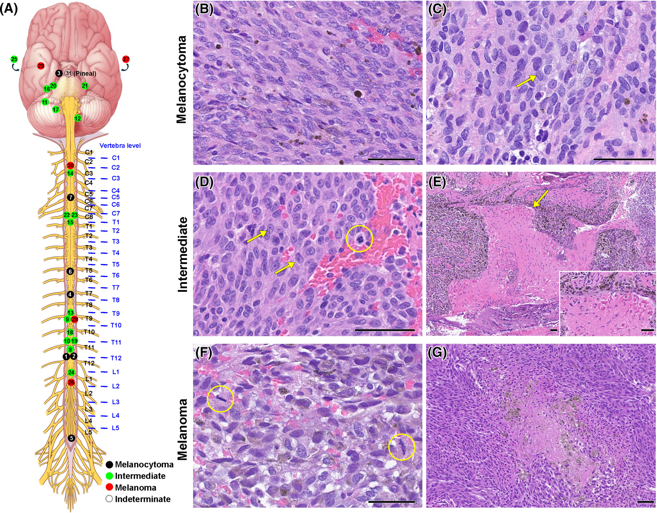
表 1. 队列临床病理与随访概览。
| 患者 | WHO 2021 诊断 | 年龄 | 性别 | 部位 | 初诊治疗 | 进展 | 生存 | ||
|---|---|---|---|---|---|---|---|---|---|
| 次数 | 首次进展时间(月) | 状态 | 死亡时间(月) | ||||||
| 1 | 黑色素细胞瘤 | 45 | 男 | T12 | STR | 4 | 33 | ADSU | |
| 2 | 黑色素细胞瘤 | 65 | 男 | T12 | Bx, RT | 1 | 11.9 | 无病生存 | |
| 3 | 黑色素细胞瘤 | 64 | 女 | R Dorello's canal | GTR, RT | 0 | 无病生存 | ||
| 4 | 黑色素细胞瘤 | 32 | 男 | T7-T8 | GTR | 7 | 51 | 死于本病 | 66.2 |
| 5 | 黑色素细胞瘤 | 33 | 女 | L5-S1 | Bx, RT | 1 | 25.1 | 死于本病 | 38.7 |
| 6 | 黑色素细胞瘤 | 51 | 女 | T5-T6 | STR, RT | 2 | 24.2 | 不详 | |
| 7 | 黑色素细胞瘤 | 50 | 男 | C5 | 不详 | 不详 | 不详 | ||
| 8 | 中间级别黑色素细胞肿瘤 | 66 | 女 | T11-T12 | Bx, RT | 0 | 带病生存 | ||
| 9 | 中间级别黑色素细胞肿瘤 | 54 | 女 | T9-T10 | STR, RT | 0 | 带病生存 | ||
| 10 | 中间级别黑色素细胞肿瘤 | 79 | 男 | T11 | STR | 1 | 22.4 | 带病生存 | |
| 11 | 中间级别黑色素细胞肿瘤 | 77 | 女 | R PF | STR | 0 | 带病生存 | ||
| 12 | 中间级别黑色素细胞肿瘤 | 53 | 女 | L foramen magnum | STR, RT | 1 | 19.3 | 带病生存 | |
| 13 | 中间级别黑色素细胞肿瘤 | 70 | 男 | T9 | STR | 2 | 1.1 | 死于本病 | 2 |
| 14 | 中间级别黑色素细胞肿瘤 | 79 | 男 | C2-3 | STR | 0 | 死于本病 | 1.6 | |
| 15 | 中间级别黑色素细胞肿瘤 | 63 | 男 | T1 | STR | 3 | 15.1 | 死于本病 | 50.5 |
| 16 | 中间级别黑色素细胞肿瘤 | 45 | 女 | R IAC to CPA | GTR | 2 | 8.2 | 死于本病 | 25.3 |
| 17 | 中间级别黑色素细胞肿瘤 | 33 | 女 | R suboccipital | GTR, RT, Immuno | 4 | 48.7 | 死于本病 | 61.7 |
| 18 | 中间级别黑色素细胞肿瘤 | 40 | 女 | T10-T11 | Bx, RT, Chemo | 2 | 36.5 | 死于本病 | 68.8 |
| 19 | 中间级别黑色素细胞肿瘤 | 37 | 女 | T11 | GTR | 4 | 29.9 | 死于本病 | 90.9 |
| 20 | 中间级别黑色素细胞肿瘤 | 27 | 女 | R CPA | STR, RT | 1 | 37.1 | 死于本病 | 68.9 |
| 21 | 中间级别黑色素细胞肿瘤 | 71 | 男 | L CPA | STR | 不详 | 不详 | ||
| 22 | 中间级别黑色素细胞肿瘤 | 36 | 男 | C6-T2 | 不详 | 不详 | 不详 | ||
| 23 | 中间级别黑色素细胞肿瘤 | 80 | 男 | C7 | 不详 | 不详 | 不详 | ||
| 24 | 中间级别黑色素细胞肿瘤 | 61 | 男 | CM | 不详 | 不详 | 不详 | ||
| 25 | 中间级别黑色素细胞肿瘤 | 76 | 男 | R parietal | 不详 | 不详 | 不详 | ||
| 26 | 黑色素瘤 | 61 | 女 | T12-S1 | STR, RT, Chemo, Immuno | 1 | 8.8 | 带病生存 | |
| 27 | 黑色素瘤 | 65 | 男 | L parietal | GTR, RT | 2 | 29.3 | 死于本病 | 30.2 |
| 28 | 黑色素瘤 | 59 | 女 | T9-T10 | GTR | 5 | 1.5 | 死于本病 | 25.3 |
| 29 | 黑色素瘤 | 51 | 女 | R temporal | Bx | 0 | 死于本病 | 0.9 | |
| 30 | 黑色素瘤 | 58 | 女 | C1-2 | GTR | 1 | 2.5 | 死于本病 | 3.1 |
| 31 | 不确定 | 82 | 女 | Pineal region | Bx, RT | 不详 | ADSU | ||
26 例行分子检测:GNAQ/GNA11 突变 21/24;BAP1 突变 5/23(IMT 2、黑色素瘤 3);EIF1AX 9/21(黑色素细胞瘤 1、IMT 8);SF3B1 3/23(IMT 2、黑色素瘤 1)。详见表 2。
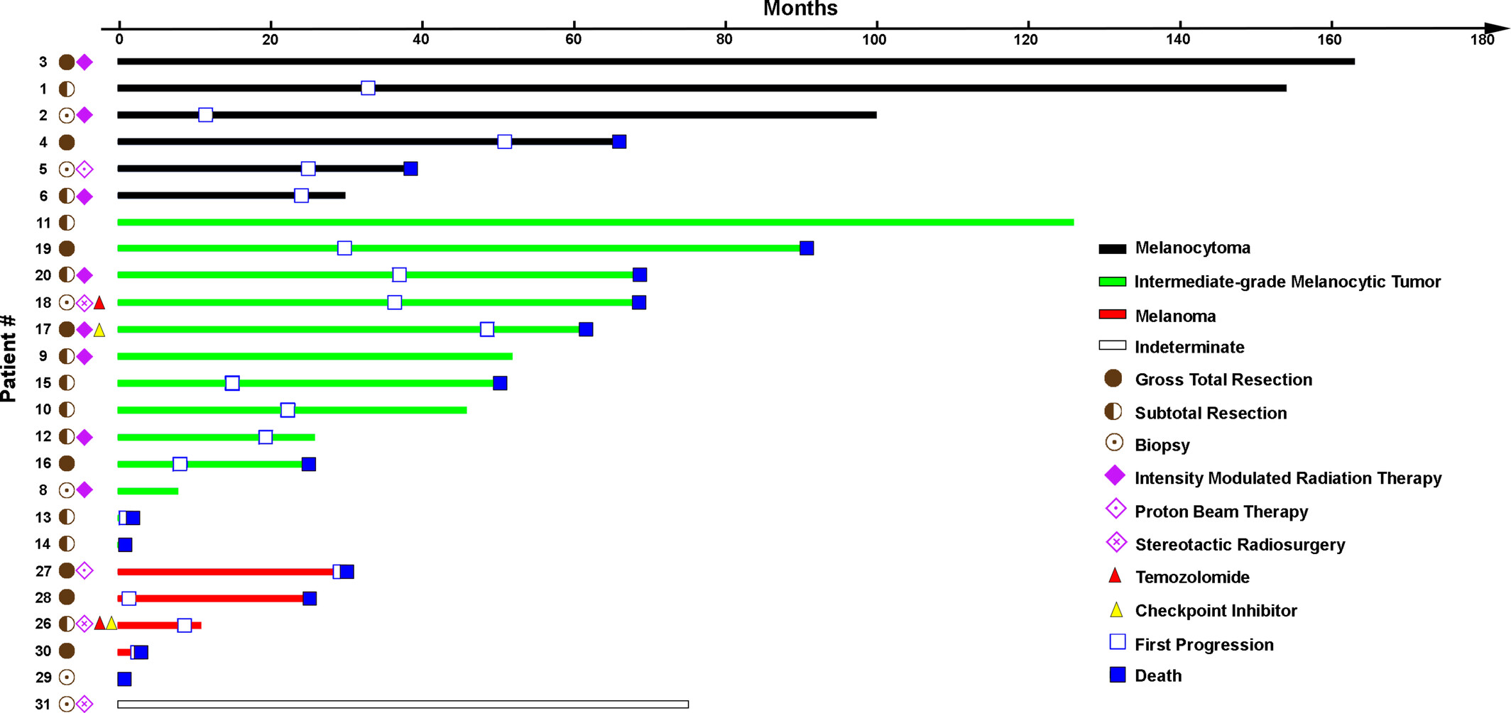
25 例有可用随访(图 2)。黑色素细胞瘤 6 例中 5 例、IMT 13 例中 9 例、黑色素瘤 5 例中 4 例出现进展,提示各级别进展频率均不低。
表 2. 分子改变谱。
| 患者 | 诊断 | GNAQ/GNA11 | BAP1 | EIF1AX | SF3B1 |
|---|---|---|---|---|---|
| 1 | 黑色素细胞瘤 | 野生型 | 未测 | 未测 | 未测 |
| 2 | 黑色素细胞瘤 | GNAQ | 野生型 | 野生型 | 野生型 |
| 5 | 黑色素细胞瘤 | GNA11 | 野生型 | 未测 | 野生型 |
| 7 | 黑色素细胞瘤 | GNAQ | 野生型 | p.G6V | 野生型 |
| 8 | 中间级别黑色素细胞肿瘤 | GNAQ | 野生型 | 野生型 | p.R625H |
| 9 | 中间级别黑色素细胞肿瘤 | GNAQ | 野生型 | 野生型 | 野生型 |
| 10 | 中间级别黑色素细胞肿瘤 | GNA11 | 野生型 | p.W70Ra | 野生型 |
| 12 | 中间级别黑色素细胞肿瘤 | GNAQ | 野生型 | p.G9R | 野生型 |
| 13 | 中间级别黑色素细胞肿瘤 | GNA11 | p.W202R | p.G6D | 野生型 |
| 14 | 中间级别黑色素细胞肿瘤 | GNAQ | 野生型 | c.17-2A>Ta | 野生型 |
| 15 | 中间级别黑色素细胞肿瘤 | GNA11 | 野生型 | 野生型 | p.G740E |
| 16 | 中间级别黑色素细胞肿瘤 | GNAQ | p.L112fs*1 | 野生型 | 野生型 |
| 17 | 中间级别黑色素细胞肿瘤 | GNAQ | 野生型 | p.K3_N4insPKa | 野生型 |
| 20 | 中间级别黑色素细胞肿瘤 | GNAQ | 野生型 | 野生型 | 野生型 |
| 21 | 中间级别黑色素细胞肿瘤 | GNAQ | 野生型 | c.17-1G>Ta | 野生型 |
| 22 | 中间级别黑色素细胞肿瘤 | GNAQ | 野生型 | 野生型 | 野生型 |
| 23 | 中间级别黑色素细胞肿瘤 | 野生型 | 野生型 | p.G8R | 野生型 |
| 24 | 中间级别黑色素细胞肿瘤 | GNAQ | 野生型 | 野生型 | 野生型 |
| 25 | 中间级别黑色素细胞肿瘤 | GNA11 | 野生型 | p.G9D | 野生型 |
| 26 | 黑色素瘤 | GNA11 | 野生型 | 未测 | 野生型 |
| 27 | 黑色素瘤 | 野生型 | p.D400Efs*2 | 野生型 | 野生型 |
| 28 | 黑色素瘤 | GNA11 | p.W196* | 野生型 | 野生型 |
| 29 | 黑色素瘤 | GNAQ | p.S497Yfs*38 | 野生型 | 野生型 |
| 30 | 黑色素瘤 | GNAQ | 野生型 | 野生型 | p.H662D |
图 2.
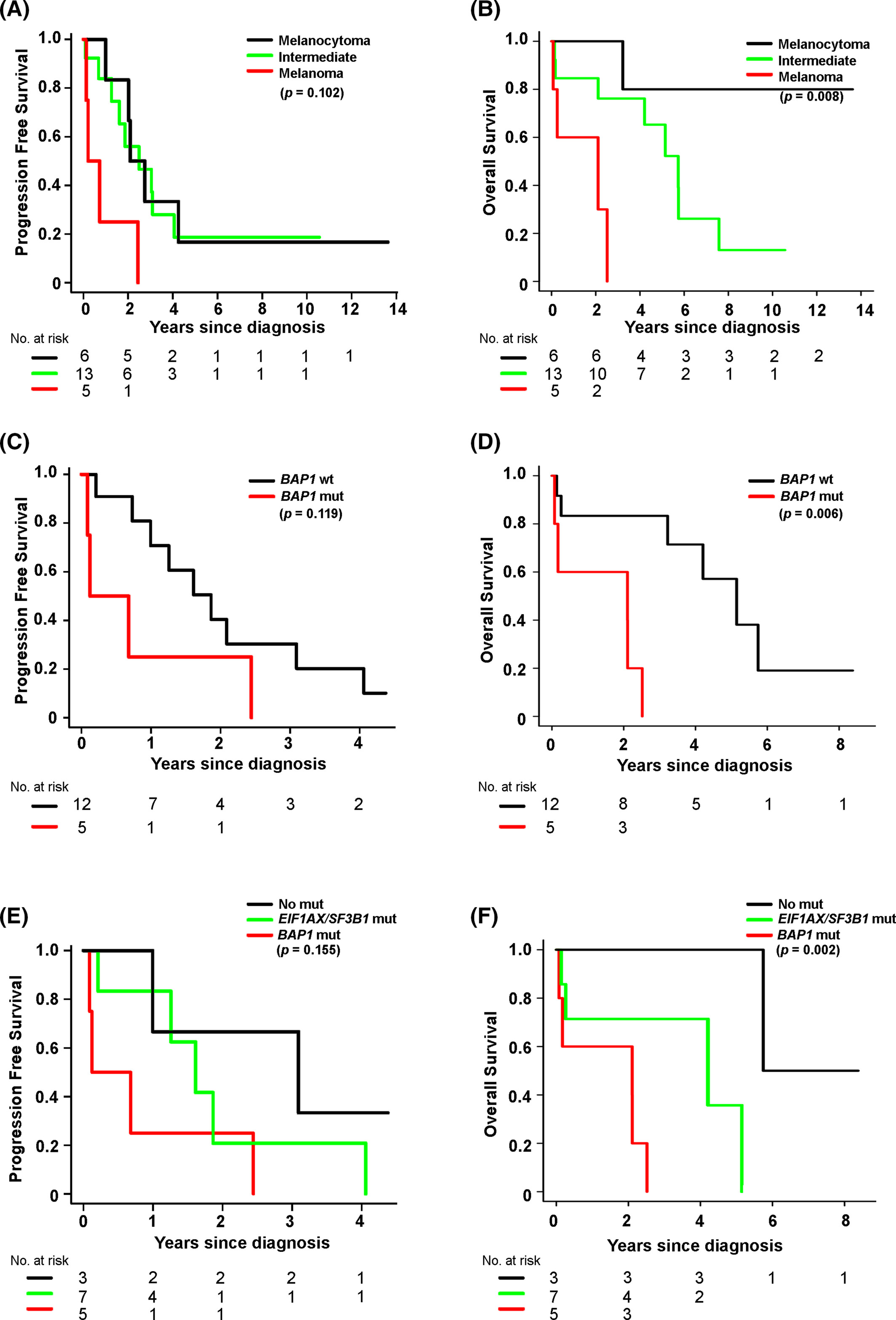
黑色素细胞瘤、IMT、黑色素瘤的 2 年 PFS 分别为 83.3%(95% CI 53.5–100)、55.9%(26.7–85.2)、25.0%(0–67.4);5 年 OS 分别为 80.0%(44.9–100)、65.3%(37.0–93.6)、0%。与黑色素细胞瘤相比,黑色素瘤 OS 显著较差(p=0.002)(图 3A,B)。
BAP1 突变与较差 OS 相关(HR 8.73,p=0.006)(图 3C,D)。按 EIF1AX/SF3B1 与 BAP1 改变组合分层的生存曲线见图 3E,F。
治疗上,多数患者接受手术±放疗;部分加化疗或免疫治疗。在本队列描述性分析中,初始最大安全切除似乎与较好结局相关,而放疗与全身治疗未显示清晰额外获益;该观察受选择偏倚与样本量限制。
IMT 组内结局异质:部分长期稳定,部分反复进展甚至死亡,提示单靠现行组织学标准不足以充分风险分层。
不确定分类的 1 例因组织有限未能可靠分级,随访状态见表 1。
图 3.
4 讨论
我们报告单中心 31 例,结合随访分析组织学、分子改变、治疗与结局的关联。结果显示各级别 CMMN 均有相当比例进展;黑色素瘤组织学及 BAP1 突变预示显著较差 OS。
IMT 组结局异质,提出仅靠组织学是否足以风险分层的问题。本组 IMT 常有 EIF1AX 等改变,而 BAP1 突变更多见于侵袭性病变,支持将分子信息纳入临床解读。
GNAQ/GNA11 高频支持与葡萄膜黑色素瘤等 Gαq 通路驱动肿瘤的生物学联系,并有助于与转移性皮肤黑色素瘤鉴别。
手术仍是主要手段;最大安全切除在部分患者中似有益。放疗与全身治疗(含替莫唑胺、免疫检查点抑制剂)在本回顾性队列中未显示一致获益,需前瞻性数据验证。
研究局限包括回顾性设计、罕见病导致的样本量小、分子检测覆盖不完全及治疗异质性。部分历史病例按当代标准重分类,可能引入时代偏倚。
尽管如此,本队列提供了 CMMN 在真实世界中的分级分布、分子谱与结局参照,并强调 BAP1 检测的潜在预后价值。
未来多中心合作有助于验证分子–组织学整合分级,并探索针对 Gαq 通路或 BAP1 相关缺陷的治疗策略。
结论:CMMNs 无论 WHO 级别均具较高复发倾向;黑色素瘤组织学与 BAP1 突变与显著较差总生存相关。最大安全切除仍是关键,分子检测尤其是 BAP1 有助于风险分层。
Abstract
Circumscribed meningeal melanocytic neoplasms (CMMNs) represent a spectrum of rare central nervous system (CNS) tumors. Knowledge of their clinical behavior and molecular correlates remains limited. In this single-institution retrospective study, we analyzed 31 patients (14 male, 17 female; median age, 59 years) diagnosed with CMMN between 2004 and 2025. In this patient cohort, based on the current World Health Organization Central Nervous System Tumor Classification (WHO CNS5) criteria, the majority of cases (18, 58.1%) fell into the intermediate-grade melanocytic tumor (IMT) category based on histology, with either increased mitotic activity, CNS invasion, or both; 7 (22.6%) were melanocytomas, 5 (16.1%) melanomas, and 1 (3.2%) indeterminate case because of limited tissue. By next-generation sequencing, mutations were identified in GNAQ/GNA11 in 21/24 cases, BAP1 in 5/23 (2 IMT, 3 melanoma), EIF1AX in 9/21 (1 melanocytoma, 8 IMT), and SF3B1 in 3/23 (2 IMT, 1 melanoma). Primary sites were spinal (20), posterior fossa (6), and supratentorial (5). During follow-up, progression was observed in 5/6 melanocytomas, 9/13 IMTs, and 4/5 melanomas, suggesting a high frequency of progression across all groups. Two-year progression-free survival was 83.3% (95% confidence interval [CI], 53.5%–100.0%) for melanocytoma, 55.9% (95% CI, 26.7%–85.2%) for IMT, and 25.0% (95% CI, 0.0%–67.4%) for melanoma. Five-year overall survival (OS) was 80.0% (95% CI, 44.9%–100.0%) for melanocytoma, 65.3% (95% CI, 37.0%–93.6%) for IMT, and 0.0% for melanoma. OS was significantly worse in melanoma compared with melanocytoma ( p = 0.002). BAP1 mutation correlated with worse OS (hazard ratio 8.73, p = 0.006). Upfront maximal safe resection appeared to be associated with improved outcomes in selected patients, whereas radiotherapy and systemic therapy did not demonstrate clear additional benefit in this cohort. In summary, our study shows that all CMMNs, irrespective of grade, have a high propensity to recur and melanoma histology and BAP1 mutation are associated with significantly worse OS.
1 INTRODUCTION
Primary melanocytic lesions are rare tumors arising from leptomeningeal melanocytes, accounting for approximately 0.03% of all central nervous system (CNS) neoplasms [ 1 ]. These tumors may arise within the spinal canal, posterior fossa, and supratentorial compartments, and can be diffuse or circumscribed [ 1 - 4 ]. Before 2021, circumscribed meningeal melanocytic neoplasms (CMMN) were histologically classified into melanocytoma and melanoma. However, diagnostically challenging intermediate lesions, with histologic features lying between these two extremes, were recognized. We have previously described an “intermediate-grade” group based on histologic features of increased mitotic activity and CNS invasion [ 5 ]. The WHO 2021 classification (5th edition) formally introduced the intermediate-grade tumor (IMT), and the histologic spectrum of CMMNs now comprises three tiers: (1) melanocytoma, characterized by limited cytologic atypia, absent or nearly absent mitotic activity, no necrosis, and no CNS invasion; (2) IMT, defined by a mitotic count of 0.5–1.5 mitoses/mm 2 and/or CNS invasion, with limited cytologic atypia and no necrosis; and (3) melanoma, defined by a mitotic count >1.5 mitoses/mm 2 and/or necrosis, often accompanied by marked cytologic atypia [ 6 ]. However, since this reclassification, no large, dedicated clinical series specifically comparing clinical outcomes across all three groups has been published.
At the molecular level, CMMNs are characterized by mutually exclusive activating mutations in GNAQ , GNA11 , PLCB4 , or CYSLTR2 , which are considered the first step in oncogenesis. Among them, GNAQ and GNA11 mutations are most common and are identified in approximately 60%–70% of cases across the histologic spectrum [ 7 - 9 ], a mutational profile similar to uveal melanoma and blue nevus [ 10 ]. Additional alterations in BAP1 , EIF1AX , and SF3B1 have also been reported in CMMNs [ 9 , 11 - 13 ]. In uveal melanoma, these secondary alterations carry well-established prognostic significance: BAP1 loss is associated with high metastatic risk and poor survival, whereas EIF1AX and SF3B1 mutations confer a relatively favorable prognosis [ 14 ]. Limited data in CMMNs suggest that alterations in BAP1 , EIF1AX , and SF3B1 may all be associated with more aggressive clinical behavior [ 9 , 12 , 13 ], but the small size of the cohorts precludes definite conclusions. The DNA methylation profile of CMMN is not distinct from that of uveal melanoma or blue nevus-like melanoma and does not reliably distinguish melanocytoma from melanoma [ 11 , 15 ] limiting its diagnostic utility. Therefore, the diagnosis and classification of CMMN remain primarily based on histopathologic features and genetic alterations.
Our study aims to characterize the clinicopathologic and molecular features of CMMNs in a single-institution cohort of 31 patients, and to correlate CNS5 WHO histologic grade and mutational profiles with clinical outcome.
2 MATERIALS AND METHODS
2.1 Patient cohort, imaging and pathological review
We identified 31 CMMN patients diagnosed and/or followed at the Mayo Clinic (2004–2025). The inclusion criteria were as follows: (1) primary CMMN arising from the leptomeninges on imaging; (2) histopathologic confirmation of a melanocytic neoplasm with a typical immunoprofile; and (3) absence of evidence of metastasis from an extracranial primary tumor. The study was approved by the Institutional Review Board, and all procedures were conducted in accordance with the ethical standards of the Declaration of Helsinki.
The available magnetic resonance (MR) images of intracranial and spinal tumors were reviewed by an experienced board-certified neuroradiologist (TJK) for lesion location (intra-axial/intramedullary vs. extra-axial) and the presence or absence of inherent pre-gadolinium T1-weighted hyperintensity. Data collected from chart review included extent of resection (gross total resection [GTR] vs. subtotal resection [STR]), adjuvant treatment (chemotherapy and/or radiation therapy [RT]), follow-up time, progression, management of progression, and outcome.
Archived hematoxylin and eosin (H&E) slides and immunohistochemical (IHC) stains were retrospectively reviewed in all cases by a neuropathologist (CG) blinded to the clinical and mutational status. The diagnostic criteria for CMMNs were based on the 2021 WHO Classification of Tumors of the Central Nervous System (CNS5). Available IHC stains included S100 ( n = 21), SOX10 ( n = 10), Melan A ( n = 18), HMB-45 ( n = 13), BRAF V600E ( n = 7), Collagen IV ( n = 17), PRKAR1A ( n = 8), and BAP1 ( n = 5).
2.2 Molecular testing
Molecular testing was performed either at diagnosis or for this study in 26 (of 31) cases. Next-generation sequencing (NGS) was performed at Mayo Clinic using a limited panel covering BRAF , GNA11 , GNAQ , KIT , and NRAS ( n = 1), or a slightly larger panel covering BAP1 , BRAF , CDKN2A , CTNNB1 , EIF1AX , GNA11 , GNAQ , HRAS , KIT , KRAS , MAP2K1 , MAP2K2 , NF1 , NRAS , SF3B1 , TERT promoter, and TP53 ( n = 18). NGS testing was also performed at other laboratories and included Tempus xT 648-gene panel ( n = 1), FoundationOne panel ( n = 1), University of Pittsburgh Medical Center (UPMC) Oncomine panel ( n = 1), Emory Expanded Cancer Mutation Profile and Interpretation panel ( n = 1), and Northwestern Memorial Hospital laboratory panel ( n = 1). One additional case underwent only BRAF V600E polymerase chain reaction (PCR)-based testing and targeted KIT (exons 11, 13, and 17) testing by PCR followed by Sanger sequencing at Mayo Clinic. One case (diagnosed as CMMN with indeterminate classification) failed testing (Table 2 ).
2.3 Statistical analysis
Patient demographic and clinicopathologic data were summarized with frequencies and percentages or medians, interquartile ranges, and ranges, as appropriate. Progression-free survival (PFS) was defined as the time from diagnosis to progression, censoring patients without progression at time of last follow-up or death. Overall survival (OS) was defined as the time from diagnosis to death from any cause (censoring patients at time of last follow-up for those still alive). PFS and OS were estimated at 2 and 5 years (respectively) following diagnosis with the Kaplan–Meier method and were compared (i.e., by mutation result) using likelihood ratio tests from Cox proportional hazards regression models. For comparisons between the three patient groups, statistical significance was defined as p < 0.017 (Bonferroni). For all other comparisons, p -values less than 0.05 were considered statistically significant. All analyses were performed using SAS version 9.4 (SAS Institute Inc., Cary, NC) or R version 4.4.1.
3 RESULTS
The cohort included 14 males and 17 females, with a median age of 59 years (range, 27–82 years). MR imaging of the tumors was available for review in 30 (of 31) subjects; for one spinal cord case (Patient 23), only an imaging report was available. Among all cases, five tumors were located supratentorially, six infratentorially, and 20 within the spinal canal (Table 1 and Figure 1A ).
Of the 19 spinal tumors with imaging available for review, 11 (57.9%) were intramedullary, 7 (36.8%) were extramedullary, and 1 (5.3%) was intramedullary with multiple intradural extramedullary lesions, presumed to represent cerebrospinal fluid (CSF) metastases. Of the 11 intracranial tumors, 8 (72.7%) were extra-axial (including one intraventricular tumor), 2 (18.2%) were intra-axial, and 1 (9.1%) was an intra-axial lesion with CSF dissemination of uncertain leptomeningeal versus peripheral intraparenchymal origin.
Many tumors were intrinsically hyperintense on pre-gadolinium T1-weighted imaging, either partially or completely, a feature associated with melanotic neoplasms. This finding was present in 13 (of 19, 68.4%) spinal tumors and 9 (of 11, 81.8%) intracranial tumors.
Figure 1.
Table 1. Clinicopathologic and follow-up overview of the cohort.
| Patient | WHO 2021 diagnosis | Age | Sex | Location | Treatment at initial diagnosis | Progression | Survival | ||
|---|---|---|---|---|---|---|---|---|---|
| No. | Time to first progression (m) | Status | Time to death (m) | ||||||
| 1 | Melanocytoma | 45 | M | T12 | STR | 4 | 33 | ADSU | |
| 2 | Melanocytoma | 65 | M | T12 | Bx, RT | 1 | 11.9 | AWOD | |
| 3 | Melanocytoma | 64 | F | R Dorello's canal | GTR, RT | 0 | AWOD | ||
| 4 | Melanocytoma | 32 | M | T7-T8 | GTR | 7 | 51 | DOD | 66.2 |
| 5 | Melanocytoma | 33 | F | L5-S1 | Bx, RT | 1 | 25.1 | DOD | 38.7 |
| 6 | Melanocytoma | 51 | F | T5-T6 | STR, RT | 2 | 24.2 | Unk | |
| 7 | Melanocytoma | 50 | M | C5 | Unk | Unk | Unk | ||
| 8 | IMT | 66 | F | T11-T12 | Bx, RT | 0 | AWD | ||
| 9 | IMT | 54 | F | T9-T10 | STR, RT | 0 | AWD | ||
| 10 | IMT | 79 | M | T11 | STR | 1 | 22.4 | AWD | |
| 11 | IMT | 77 | F | R PF | STR | 0 | AWD | ||
| 12 | IMT | 53 | F | L foramen magnum | STR, RT | 1 | 19.3 | AWD | |
| 13 | IMT | 70 | M | T9 | STR | 2 | 1.1 | DOD | 2 |
| 14 | IMT | 79 | M | C2-3 | STR | 0 | DOD | 1.6 | |
| 15 | IMT | 63 | M | T1 | STR | 3 | 15.1 | DOD | 50.5 |
| 16 | IMT | 45 | F | R IAC to CPA | GTR | 2 | 8.2 | DOD | 25.3 |
| 17 | IMT | 33 | F | R suboccipital | GTR, RT, Immuno | 4 | 48.7 | DOD | 61.7 |
| 18 | IMT | 40 | F | T10-T11 | Bx, RT, Chemo | 2 | 36.5 | DOD | 68.8 |
| 19 | IMT | 37 | F | T11 | GTR | 4 | 29.9 | DOD | 90.9 |
| 20 | IMT | 27 | F | R CPA | STR, RT | 1 | 37.1 | DOD | 68.9 |
| 21 | IMT | 71 | M | L CPA | STR | Unk | Unk | ||
| 22 | IMT | 36 | M | C6-T2 | Unk | Unk | Unk | ||
| 23 | IMT | 80 | M | C7 | Unk | Unk | Unk | ||
| 24 | IMT | 61 | M | CM | Unk | Unk | Unk | ||
| 25 | IMT | 76 | M | R parietal | Unk | Unk | Unk | ||
| 26 | Melanoma | 61 | F | T12-S1 | STR, RT, Chemo, Immuno | 1 | 8.8 | AWD | |
| 27 | Melanoma | 65 | M | L parietal | GTR, RT | 2 | 29.3 | DOD | 30.2 |
| 28 | Melanoma | 59 | F | T9-T10 | GTR | 5 | 1.5 | DOD | 25.3 |
| 29 | Melanoma | 51 | F | R temporal | Bx | 0 | DOD | 0.9 | |
| 30 | Melanoma | 58 | F | C1-2 | GTR | 1 | 2.5 | DOD | 3.1 |
| 31 | Indeterminate | 82 | F | Pineal region | Bx, RT | Unk | ADSU | ||
Based on the CNS5 criteria, the tumors were classified as melanocytoma ( n = 7), IMT ( n = 18), and melanoma ( n = 5). One case remained indeterminate because insufficient tissue was available for definitive histological characterization (Table 1 ). Morphologically, melanocytomas exhibited a bland spindle cell architecture (Figure 1B,C ), frequently with prominent nuclear grooves (Figure 1C , arrow), lacked mitotic activity, and had a low Ki-67 labeling index (0%–2%). IMTs typically demonstrated spindle to epithelioid morphology, occasionally with cytologic atypia with prominent nucleoli (Figure 1D , arrows), increased mitotic activity (Figure 1D , circle, median 1 per 10 high-power fields (HPF), range 0–3, or 0.5 mitoses/mm 2 ), and a Ki-67 labeling index ranging from 0% to 7%. Parenchymal/CNS invasion was observed in 8 (of 18, 44.4%) IMTs, and the tumor interface often showed reactive changes, including the presence of Rosenthal fibers (Figure 1E , arrow and inset). Two (of 18, 11.1%) IMT cases showed both increased mitotic activity and CNS invasion. Melanomas displayed marked nuclear atypia, high mitotic activity (Figure 1F , circles, median 5 per 10 HPF, range 5–9, or 2.5 mitoses/mm 2 ), and frequent tumor necrosis (four of five cases, 80.0%, Figure 1G ).
By IHC, S100 was positive in tumor cells in 16 (of 21, 76.2%) cases. In contrast, SOX10 (10/10), Melan A (18/18), and HMB45 (13/13) were positive in all tested cases. Two (of five) cases showed BAP1 loss. All tested cases were negative for BRAF V600E (7/7). Expression of PRKAR1A, which is typically lost in malignant melanotic nerve sheath tumor (MMNST), was retained in eight (of eight) cases tested [ 16 ]. Pericellular collagen IV deposition, also a feature of MMNST, was absent in 17 (of 17) cases tested [ 6 ].
Molecular testing data are summarized in Table 2 . GNAQ (p.Q209L, n = 12; p.Q209P, n = 2) or GNA11 (p.Q209L, n = 7) mutations were present in 21 (of 24, 87.5%) cases, including melanocytoma (3/4, 75.0%), IMTs (14/15, 93.3%), and melanomas (4/5, 80.0%). BAP1 mutations were identified in 5 (of 23, 21.7%) cases, comprising 2 (of 15, 13.3%) IMTs and 3 (of 5, 60.0%) melanomas, which includes the two cases with BAP1 IHC loss. EIF1AX alterations were detected in 9 (of 21, 42.9%) cases, including 1 (of 2, 50.0%) melanocytomas and 8 (of 15, 53.3%) IMTs. SF3B1 mutations were identified in 3 (of 23, 13.0%) cases, including 2 (of 15, 13.3%) IMTs and 1 (of 5, 20%) melanoma. Two cases lacking a GNAQ / GNA11 mutation tested for other genes, had either an EIF1AX or a BAP1 mutation. Notably, BAP1 , EIF1AX and SF3B1 mutations were mutually exclusive, except for a single patient diagnosed with IMT that harbored a GNA11 mutation as well as both BAP1 and EIF1AX mutations.
Table 2. Molecular alteration profile.
| Patient | Diagnosis | GNAQ/GNA11 | BAP1 | EIF1AX | SF3B1 |
|---|---|---|---|---|---|
| 1 | Melanocytoma | wt | ND | ND | ND |
| 2 | Melanocytoma | GNAQ | wt | wt | wt |
| 5 | Melanocytoma | GNA11 | wt | ND | wt |
| 7 | Melanocytoma | GNAQ | wt | p.G6V | wt |
| 8 | IMT | GNAQ | wt | wt | p.R625H |
| 9 | IMT | GNAQ | wt | wt | wt |
| 10 | IMT | GNA11 | wt | p.W70Ra | wt |
| 12 | IMT | GNAQ | wt | p.G9R | wt |
| 13 | IMT | GNA11 | p.W202R | p.G6D | wt |
| 14 | IMT | GNAQ | wt | c.17-2A>Ta | wt |
| 15 | IMT | GNA11 | wt | wt | p.G740E |
| 16 | IMT | GNAQ | p.L112fs*1 | wt | wt |
| 17 | IMT | GNAQ | wt | p.K3_N4insPKa | wt |
| 20 | IMT | GNAQ | wt | wt | wt |
| 21 | IMT | GNAQ | wt | c.17-1G>Ta | wt |
| 22 | IMT | GNAQ | wt | wt | wt |
| 23 | IMT | wt | wt | p.G8R | wt |
| 24 | IMT | GNAQ | wt | wt | wt |
| 25 | IMT | GNA11 | wt | p.G9D | wt |
| 26 | Melanoma | GNA11 | wt | ND | wt |
| 27 | Melanoma | wt | p.D400Efs*2 | wt | wt |
| 28 | Melanoma | GNA11 | p.W196* | wt | wt |
| 29 | Melanoma | GNAQ | p.S497Yfs*38 | wt | wt |
| 30 | Melanoma | GNAQ | wt | wt | p.H662D |
Figure 2.
The clinical outcomes are listed in Table 1 , Figures 2 and 3 . Among 31 patients, 25 had available follow-up data (median follow-up: 30.2 months), including 6 with melanocytoma, 13 with IMT, 5 with melanoma, and 1 with indeterminate tumor. In the melanocytoma group (median follow-up: 66.2 months), five (of six) patients experienced progression (range, 12–51 months), and two patients died of disease; the 2-year PFS was 83.3% (95% confidence interval [CI], 53.5%–100.0%), and the 5-year OS was 80.0% (95% CI, 44.9%–100.0%). In the IMT group (median follow-up: 26.4 months), 9 (of 13) patients experienced progression (range, 1–49 months), and 8 patients died of disease; the 2-year PFS was 55.9% (95% CI, 26.7%–85.2%), and the 5-year OS was 65.3% (95% CI, 37.0%–93.6%). In the melanoma group (median follow-up: 11.1 months), four (of five) patients experienced progression (range, 2–29 months); among those four with progression, three patients died and one remained alive with disease at 11 months of follow-up; the remaining patient had leptomeningeal dissemination at diagnosis, transitioned to hospice care following biopsy, and died 1 month later. The overall 2-year PFS was 25.0% (95% CI, 0.0%–67.4%), and the 5-year OS was 0.0%. Based on a lower threshold for significance to account for three pairwise comparisons between the groups ( p < 0.017), OS was significantly worse for melanoma patients as compared with those with melanocytoma ( p = 0.002) (Figure 3A,B ). Regarding tumor location (spinal, infratentorial, and supratentorial), we found no statistically significant association between tumor location and either PFS ( p = 0.31) or OS ( p = 0.12). The number of cases was insufficient to support meaningful comparisons of PFS and OS across the three tumor locations within individual diagnostic categories.
Regardless of histologic classification, BAP1 mutation was significantly associated with worse OS (hazard ratio 8.73, p = 0.006), while no difference in PFS was observed compared with BAP1 wildtype (Figure 3C,D ). Notably, the two IMT cases with BAP1 mutations died of disease at 2 and 25 months after diagnosis, corresponding to a 5-year OS of 0.0% compared with a 5-year OS of 65.6% in the remaining BAP1 -wildtype IMTs. Significant differences in OS were observed in the overall comparison across patients without secondary mutations, those harboring EIF1AX/SF3B1 , and those with BAP1 mutations ( p = 0.002). No significant differences in PFS were observed across these three groups (Figure 3E,F ).
The initial surgical management and adjuvant therapies received are summarized in Table 1 and Figure 2 . At initial diagnosis, six patients underwent biopsy, 11 underwent STR, and eight underwent GTR. Adjuvant RT was administered to 13 patients as follows: 5 (Patients 2, 3, 8, 12, 20) received photon RT to a total dose of 50–60 Gy, 4 (Patients 6, 9, 17, and 18) received photon RT to 20–49 Gy, 1 (Patient 27) received proton RT to 60 Gy, 1 (Patient 5) received proton craniospinal irradiation (CSI) for disseminated disease, 1 (Patient 31) received Gamma Knife radiosurgery alone, and 1 (Patient 26) received multimodality RT consisting of Gamma Knife to a cerebellar lesion, photon RT to 20 Gy to a separate brain lesion, and photon RT to 27 Gy to each of two spinal sites. Three patients (Patients 17, 18, and 26) additionally received systemic therapy: one received temozolomide for 1 month followed by ipilimumab/nivolumab for two cycles, both of which were discontinued because of toxicity; one received temozolomide for 10–11 months before discontinuation because of toxicity; and one received three doses of ipilimumab, which was also stopped because of toxicity. Twelve patients did not receive any adjuvant therapy.
Following initial diagnosis, three patients (Patient 14, 29, and 30) experienced rapid disease progression culminating in death before initiation of adjuvant therapy. Eighteen patients developed documented progression. At first progression, treatments were as follows: one underwent GTR alone; five received RT alone (one Gamma Knife, one stereotactic radiosurgery [SRS] to two cerebellar lesions plus 45 Gy to a thoracic lesion, two received 40–50.4 Gy, and one received proton CSI); four received checkpoint inhibitor therapy alone (nivolumab and/or ipilimumab); one received rapamycin but was transitioned to temozolomide because of toxicity; one underwent STR plus RT to 50.4 Gy; one underwent surgery plus temozolomide; and one received RT to two sites (30–40 Gy) plus checkpoint inhibitor therapy (ipilimumab/nivolumab). One patient elected observation alone. Details of treatment following first progression were unclear for two patients. One patient died of disease too quickly following first progression to allow for treatment. Four patients did not experience progression: these patients had undergone STR alone, biopsy with 50.4 Gy adjuvant RT, STR with 45 Gy adjuvant RT, and GTR with 54 Gy adjuvant RT, respectively.
Figure 3.
4 DISCUSSION
We present 31 cases from a single institution with clinical follow-up and analyze the correlations between histologic features, molecular alterations, clinical management, and outcomes. In our patient cohort based on the current WHO CNS5 classification criteria for CMMN, the majority of cases (18/31, 58.1%) fell into the IMT category based on histology, with either increased mitotic activity or CNS invasion or both. All groups had a high frequency of progression without significant difference among the three groups; however, OS differed significantly among the groups. Specifically, with a lower threshold for significance ( p < 0.017) based on three pairwise comparisons between the groups, OS was significantly worse for melanoma patients as compared with those with melanocytoma. The IMT group showed a suggestive trend toward intermediate OS, worse than melanocytoma but better than melanoma. These findings support the clinical relevance of the IMT category, which appears to capture the histologic and prognostic heterogeneity within the CMMN spectrum.
The heterogeneous outcomes observed within the IMT group raise the question of whether histologic features alone are sufficient for risk stratification. In our cohort, IMTs frequently harbored additional alterations, including BAP1 , EIF1AX , and SF3B1 . We also showed that the two IMT patients with BAP1 mutations had a 5-year OS of 0.0%, suggesting that the presence of a BAP1 mutation may define a high-risk molecular subset even in IMTs that do not meet histologic criteria for melanoma.
In our cohort, among all CMMN cases, BAP1 mutations were present in IMT and melanoma and were associated with significantly worse OS. This is consistent with prior studies demonstrating that BAP1 loss is associated with aggressive behavior and poor prognosis in CNS melanocytic neoplasms [ 9 , 11 ]. By contrast, no significant difference in OS was observed for the presence of EIF1AX or SF3B1 alterations, which may be attributable to the small sample size and limited statistical power. However, these alterations were generally absent in melanocytomas and were more frequently observed in IMTs and melanomas in our cohort, supporting their association with higher histologic grade. Larger multi-institutional studies are needed to clarify the prognostic significance of these alterations in CMMN.
Reported outcomes for CMMN vary widely across studies and are strongly influenced by extent of resection and use of adjuvant therapy [ 4 , 17 , 18 ]. In our cohort, overall treatment exposure prior to disease progression was modest. Upfront surgical management was widely implemented, and most patients, both those who progressed and those who did not, underwent at least STR at initial presentation. A subset of patients who underwent upfront resection, including both GTR and STR, appeared to experience longer time to progression and improved OS. However, outcomes were heterogeneous: some patients who underwent extensive upfront surgery progressed rapidly, whereas others treated with biopsy alone demonstrated more favorable clinical courses. While this suggests that intrinsic tumor biology may play an important role in outcomes, our data also support a potential benefit of maximal safe resection when feasible.
Upfront RT was not consistently associated with improved outcomes across the cohort. However, among patients initially managed with biopsy alone, the addition of RT appeared to yield outcomes comparable to those seen in patients treated with more extensive surgical approaches. Adjuvant RT at initial diagnosis did not appear to provide a clear benefit in patients who had undergone GTR or STR. Nevertheless, consistent with prior studies, radiotherapy may provide durable local control with minimal morbidity in surgically inaccessible tumors [ 17 , 19 ]. The use of systemic therapy, including chemotherapy and/or immunotherapy, was limited in this cohort, and patients who received these treatments upfront did not demonstrate clear evidence of improved outcomes. Although limited by sample size, these findings do not support the routine use of upfront systemic therapy.
It is worth noting that, compared with our prior CMMN cohort [ 5 ], the current cohort shows a higher recurrence/progression rate across diagnostic categories (melanocytoma, IMT, and melanoma), which may explain the lack of a statistically significant difference in PFS. The two cohorts show no significant differences in case-selection criteria, definition of recurrence, use of RT, or follow-up duration (median, 30.2 vs. 36 months in the current and prior cohorts, respectively). In the current cohort, however, biopsy-only cases are more common overall (there were no biopsy-only cases in the prior cohort), although no significant differences are observed within specific diagnostic categories (melanocytoma, IMT, and melanoma). While this finding raises the possibility that extent of resection may contribute, at least in part, to the observed differences, it should be interpreted with caution given the small sample size and the significant difference in the distribution of diagnostic categories between the current and prior cohorts (the prior cohort included only three IMT cases). It is plausible that intrinsic tumor aggressiveness and underlying tumor biology, rather than differences in treatment response alone, may play a more important role in determining clinical outcomes.
This study is limited by the rarity of the disease and the resulting small sample size, despite representing one of the largest CMMN cohorts to date. Incomplete longitudinal follow-up in a proportion of patients (particularly for the relatively small melanocytoma subset) further restricts statistical power for subgroup analyses. Additionally, the retrospective single-institution design introduces the potential for selection and referral bias. We also did not evaluate chromosomal copy number variations in the CMMN cases because of limited available material, although prior studies have shown that chromosomal copy number alterations also correlate with prognosis in CMMNs [ 11 ]. These limitations underscore the need for larger multi-institutional studies incorporating standardized molecular profiling, integrated copy-number analysis, and longitudinal clinical follow-up to further clarify the prognostic significance and clinical utility of molecular alterations in CMMNs.
In conclusion, we presented 31 CMMN cases diagnosed according to the current WHO CNS5 criteria. We showed that CMMNs have a high rate of recurrence across all three diagnostic categories but differ significantly in OS. The IMT category demonstrated an intermediate OS between melanocytoma and melanoma, supporting its clinical relevance as a distinct diagnostic category. Consistent with prior studies implicating secondary alterations in aggressive clinical behavior [ 9 , 12 ], we demonstrate a significant association between BAP1 mutation and OS. Thus, our study provides further evidence that integrated molecular profiling may complement histologic grading in the risk assessment of CMMN.