- 新增两例 SMARCB1 突变型低级别弥漫浸润性肿瘤(LGDIT),均有横纹肌样形态与 INI1 完全缺失。
- 纯合 SMARCB1 缺失,甲基化谱与既往 LGDIT 聚类一致,支持其作为独立肿瘤类型。
- 本页为 PMC OA 全文中英双语(PMC13547991)。
正文
近期研究报道了极罕见的 SMARCB1 突变型低级别弥漫浸润性肿瘤(LGDIT),扩展了中枢神经系统 SMARCB1 缺陷肿瘤的疾病谱[1–3]。与侵袭性非典型畸胎样/横纹肌样肿瘤(ATRT)相比,LGDIT 临床过程较缓和,但已有恶性转化为 ATRT 的报道;两者 DNA 甲基化谱相似,尤其接近 ATRT-MYC 亚组[1,4]。本文报告两例形态、免疫表型、分子和表观遗传特征均符合 LGDIT 的病例,并依据 cIMPACT-NOW update 10 标准讨论其能否作为 WHO 中枢神经系统肿瘤分类中的暂定类型[5]。
病例 1
患者为 18 岁男性,因注意力及行为障碍入院。MRI 示双额叶 FLAIR 高信号病变,以右额叶为主,钆增强不均匀(图 1);CT 高密度提示钙化。活检为中等细胞密度、弥漫浸润性肿瘤,背景黏液样并伴胶原。肿瘤由梭形细胞和呈横纹肌样或少突胶质细胞样的小圆细胞构成,核延长或圆形,中度异型。病灶中散在反应性星形细胞和变性神经元。未见蜂窝状外观、微血管增生或坏死。核分裂活性低,为 2 个/3 mm²。
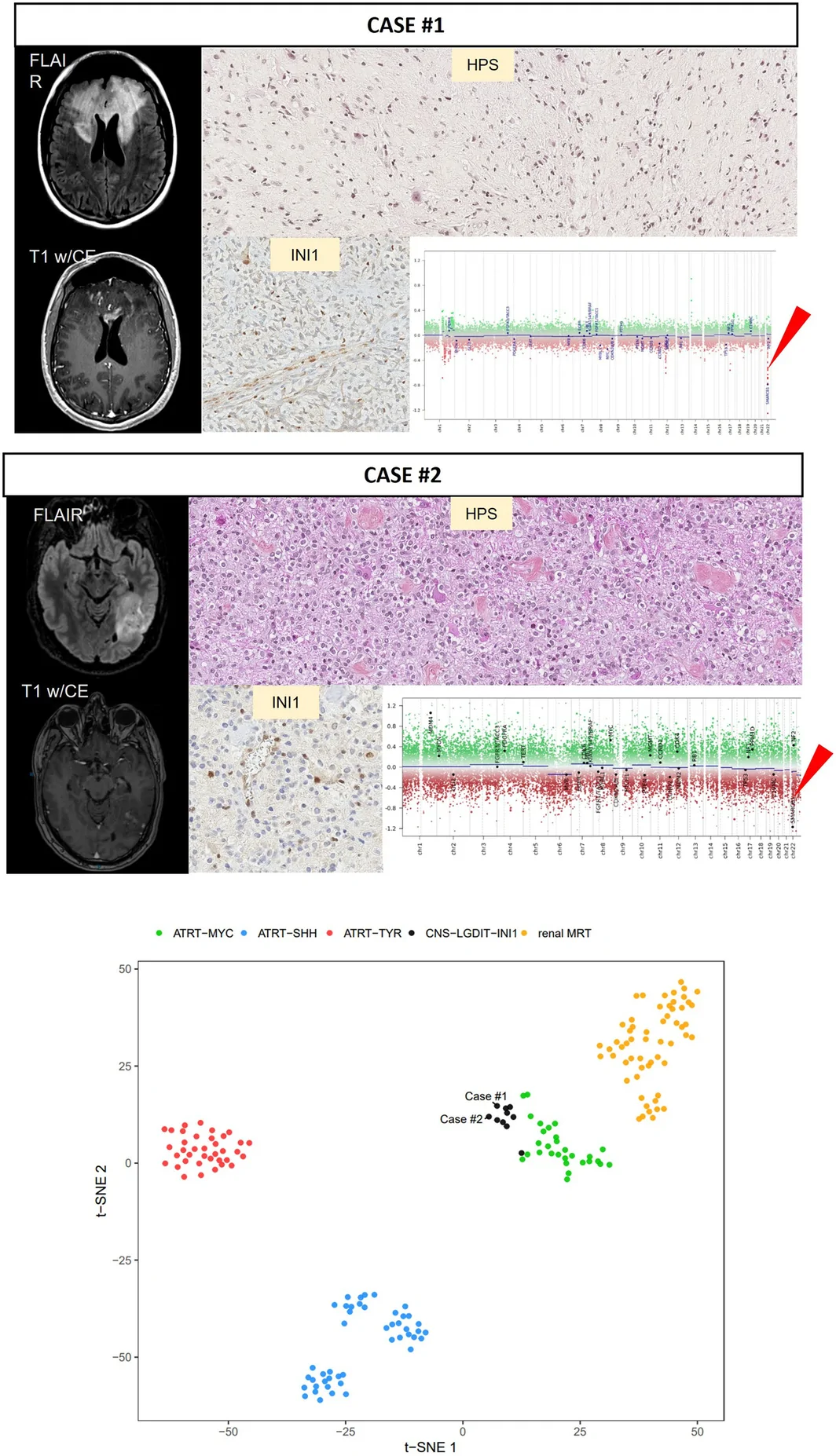
免疫组化(图 S2)示 GFAP、OLIG2、myogenin 阴性,部分细胞有血管外 CD34 表达;MIB1 标记指数约 5%。IDH1 R132H 和 H3K27M 突变蛋白不表达,无 p53 核积聚,ATRX 和 H3K27me3 核表达保留。甲基化数据的 CNV 分析提示 SMARCB1 纯合缺失,全外显子测序(WES)确认,并与肿瘤细胞 SMARCB1/INI1 核表达缺失相符(图 1)。WES 仅检出少量意义未明变异(VUS):MAPK1 c.655G>A p.(Ala219Thr),伴杂合性缺失(LOH),变异等位基因频率(VAF)41%;KMT2D c.7270C>T p.(Pro2424Ser),伴 LOH,VAF 40%;CDKN2C c.263C>T p.(Thr88Ile),VAF 21%。未检出融合转录本(Arriba 2.0.0、Fusion Catcher 1.20、StarFusion 1.9.1)。
海德堡分类器 v12.8 提示 ATRT 甲基化类别(校准分数 0.86),亚类为 ATRT-MYC(0.58);t-SNE 分析与既往 LGDIT 病例聚类(图 1),支持 LGDIT 诊断。给予卡铂/依托泊苷/环磷酰胺辅助化疗 2 个月,随后局部放疗 54 Gy(图 S1)。26 个月随访时临床稳定,但影像较术后 14 个月缓慢进展;患者最终于随访 31 个月因进展死亡。
病例 2
患者为 50 岁男性,因癫痫入院。MRI 示左顶叶轴内肿瘤,T1 增强不均匀,T2 FLAIR 高信号(图 1)。经神经心理评估后行清醒手术,次全切除。组织学见中等细胞密度的弥漫浸润性肿瘤,累及皮质和近皮质白质,背景为黏液及胶原,散在反应性星形细胞和营养不良性神经元。肿瘤细胞中小型,少突胶质样或横纹肌样,中度异型;核分裂最多 2 个/2 mm²,无微血管增生或坏死。
肿瘤细胞 GFAP、OLIG2、突触素、嗜铬粒蛋白 A、MAP2、NeuN、广谱细胞角蛋白、CD45、平滑肌肌动蛋白、desmin、myogenin、HMB45、SOX10、CD163 和 CD1a 均阴性;仅部分细胞表达 vimentin 和 CD34(图 S2)。IDH1 R132H、H3G34R 和 H3K27M 突变蛋白不表达,ATRX 和 H3K27me3 核表达保留。肿瘤细胞 INI1 核表达完全缺失(图 1),符合 SMARCB1 纯合缺失,虽样本质量欠佳,仍经 FISH、NGS 和甲基化 CNV 数据确认。NGS 另检出 KIT VUS c.1588G>A p.(Val530Ile),VAF 49%。未检出融合转录本。海德堡 v12.8 未给出可信类别,可能与样本质量有关,但 t-SNE 与既往 LGDIT 聚类,支持诊断。未予辅助抗肿瘤治疗;24 个月随访时临床、影像均稳定,MRI 残留肿瘤未变。
图 1.
综合分析与肿瘤分类
包括本文两例在内,目前六篇独立研究共报道 15 例 LGDIT(补充表 1)[1–3,6–8]。汇总资料显示其较一致的模式:主要见于青少年和年轻成人,中位年龄 19 岁(7–50 岁),男性明显占优,以幕上病变为主,癫痫是常见首发症状。但临床过程从长期稳定到 3 年内死亡不等,治疗差异很大[2,3]。甲基化分析一贯将 LGDIT 置于独立聚类,邻近 ATRT-MYC 及 SMARCB1 突变型促纤维增生性黏液样肿瘤(DMT)[9],反映 SWI/SNF 复合体缺陷及起源细胞对甲基化谱的主导影响[1,2]。转录组研究可进一步支持这种区分,如 ATRT 分子亚组已有的发现;但聚类差异也可能源于样本肿瘤含量较低或非肿瘤细胞混入。
分子层面,迄今 100% 病例均有 SMARCB1 纯合缺失,似为其单一遗传标志。相比之下,ATRT、DMT 等 INI1 缺陷肿瘤可有更广泛的变异类型,包括单核苷酸变异或小片段插入/缺失。曾报告其他 VUS 或意义不明的小缺失,但无复发性,意义尚不清楚[1–3]。虽然测序手段有限,尚无其他致病变异报道。LGDIT 不同于脑膜血管瘤病(MAM)或胶质神经元肿瘤继发的 ATRT;后者通常为边界清楚并携带 BRAF 改变的病变[10–12],这两个特点均不见于 LGDIT。
各组病例显示,LGDIT 为中到高细胞密度、常边界不清的弥漫浸润性病变,单一的横纹肌样或少突胶质细胞样细胞分布在黏液—胶原基质中。核分裂通常较少,但 Zhao 等的病例系列可达 5 个/10 高倍视野,Ki-67 指数 15%[7];无明确微血管增生或坏死。免疫表型为缺乏胶质谱系标志 GFAP、OLIG2,局灶表达 CD34 和 vimentin,INI1 核染色完全缺失。单个特征均不特异,但组合具有辨识度及可重复性。最大系列仅 6 例,世界各地的病例报道既反映罕见,也提示可能漏诊。我们依据 cIMPACT-NOW update 10 对新 CNS 肿瘤类型的标准逐项评估(表 1)[5]。
表 1. TABLE 1
| cIMPACT‐NOW criteria | Argument | Assessment |
|---|---|---|
| 1—Clinical phenotype | The clinical phenotype is partly consistent, but outcome and treatment response remain markedly heterogeneous. | Partially met |
| 2—Large‐scale molecular profiling | DNA methylation profiling places LGDIT in a separate cluster, although the available reference cohorts remain small. | Not yet fully met |
| 3—Associated molecular profile | Homozygous SMARCB1 deletion is the defining alteration, but no consistent secondary genetic event separates LGDIT from other SMARCB1‐deficient tumour types. | Not independently met |
| 4—Microscopic and immunohistochemical features | The histological and immunophenotypic features are not specific but the combination is distinctive and reproducible. | Met |
| 5—Independent publications | Although the published series remain small, LGDIT has now been reported across multiple independent studies. | Supports consideration as a provisional tumour type under the exceptional clause for very rare entities |
| 6—cIMPACT‐NOW committee approval | Not yet applicable. | Not yet applicable |
对于 LGDIT 的分类位置,可提出两种解释。其一是 ATRT-MYC 的形态变型,依据为一致的 SMARCB1 纯合缺失、横纹肌样形态以及部分复发病例进展为 ATRT;反对依据为不同的年龄分布、浸润性生长和初期缓和病程。另一种解释是:LGDIT 之于 ATRT-MYC,类似筛状神经上皮肿瘤(CRINET)之于 ATRT-TYR,即同一表观遗传谱系中的低级别、缓和对应类型。需更多整合组织学、分子与纵向随访数据,才能确定它是应正式认可的新类型,还是具有潜在恶性演变能力、极罕见的 ATRT-MYC 低级别变型。
既往治疗从单纯观察,到借用 ATRT 方案的辅助化疗和局部放疗不等。很多患者可稳定较长时间,但 15 例中有 4 例死亡,提示恶性进展风险及平衡治疗不足与过度治疗的困难。综合文献及本组两例,作者支持在可行时最大安全切除、密切影像随访,并在进展时升级治疗,通常参考 ATRT 方案,同时避免初期缓和阶段过度治疗。
作者贡献与声明
AM、GAS 起草稿件并设计图;PV 监督研究;MB、JMP、RS 完成分子分析;JP、AM、PL、LT、ABS 收集临床资料;AM、GAS、AV、ATE、PV 复核组织学。全体作者批判性修改并批准最终稿。资助及伦理声明无可报告事项。巴黎 GHU Psychiatrie et Neurosciences 和里昂 Hospices Civils 患者提供书面知情同意或不反对参加研究的声明。作者声明无利益冲突。
补充材料
图 S1 展示两例临床及影像过程:病例 1 经活检、化疗及放疗后,26 个月内影像缓慢进展而无临床症状,31 个月死亡;病例 2 清醒功能定位手术后残留小片 FLAIR 高信号、无强化灶,抗癫痫药控制良好,24 个月稳定。图 S2 为 GFAP、OLIG2、CD34、Ki-67 等免疫染色,病例 2 另有 vimentin 阳性;其 FISH 采用 ZytoLight SPEC SMARCB1 双色探针,显示 SMARCB1(22q11,绿)缺失,以 KREMEN1(22q12,红)作对照。表 S1 汇总已报道病例。文件入口见下方。
Abstract
Two new cases of low‐grade diffusely infiltrative tumour (LGDIT), SMARCB1 mutant, are described in an 18‐year‐old and a 50‐year‐old male, both with supratentorial lesions, characteristic rhabdoid histology on a myxoid‐collagenous background, and complete INI1 loss. Both tumours showed homozygous SMARCB1 deletion and clustered with previously reported LGDIT on t‐SNE analysis, in proximity to ATRT‐MYC. These observations reinforce the distinct clinicopathological profile of LGDIT and support its consideration as a provisional CNS tumour type.
Recent studies have reported very rare, low‐grade, diffusely infiltrative tumours (LGDIT), SMARCB1‐mutant, expanding the spectrum of central nervous system (CNS) SMARCB1‐deficient tumours [1, 2, 3]. Despite their indolent clinical behaviour compared with aggressive atypical teratoid and rhabdoid tumours (ATRTs), malignant transformation of LGDIT into ATRT has been described and both share a similar DNA‐methylation profile, particularly close to the ATRT‐MYC subgroup [1, 4]. Here, two cases are reported whose morphological, immunophenotypic, molecular and epigenetic profiles were consistent with LGDIT, and a potential qualification as a provisional tumour type for the World Health Organization (WHO) classification of CNS tumours is discussed, according to cIMPACT‐NOW update 10 criteria [5].
The first patient was an 18‐year‐old male admitted for attentional and behavioural deficits. Brain MRI revealed a hyperintense bifrontal lesion on FLAIR imaging, with heterogeneous gadolinium enhancement, predominantly in the right frontal lobe (Figure 1). Hyperdensities on the computed tomography suggested calcification. The biopsy showed a moderately cellular, diffusely infiltrative tumour on a myxoid and collagenous background. The tumour was composed of spindle‐shaped cells and small round cells with a rhabdoid or oligo‐like appearance, with elongated or rounded nuclei and moderate atypia. Reactive astrocytes and degenerative neurons were scattered throughout the lesion. No honeycomb appearance, microvascular proliferation or necrosis was observed. Mitotic activity was low with 2 mitoses per 3 mm2. On immunohistochemistry (Figure S2), tumour cells were negative for GFAP, OLIG2 and myogenin. A subset of cells showed CD34 extravascular expression. MIB1 labelling index was around 5%. There was no expression of IDH1 R132H or H3K27M mutant proteins, no nuclear p53 accumulation and nuclear expression of ATRX and H3K27me3 was retained. Copy number variation (CNV) analysis from DNA‐methylation data showed a homozygous SMARCB1 deletion, confirmed by whole‐exome sequencing (WES), which correlated with loss of nuclear SMARCB1/INI1 expression in the tumour cells (Figure 1). WES identified only a few variants of unknown significance (VUS): MAPK1 with loss of heterozygosity (LOH) c.655G > A p.(Ala219Thr) with a variant allele frequency (VAF) of 41%; KMT2D with LOH c.7270C > T p.(Pro2424Ser) with a VAF of 40%; CDKN2C c.263C > T p.(Thr88Ile) with a VAF of 21%. No fusion transcripts were detected (Arriba version 2.0.0; Fusion Catcher version 1.20; StarFusion version 1.9.1). DNA methylation profiling, using the Heidelberg classifier v12.8, suggested the ATRT methylation class (calibrated score 0.86) and the subclass of ATRT‐MYC (calibrated score 0.58), and the t‐SNE analysis showed clustering with previously described cases of LGDIT (Figure 1). These findings supported the diagnosis of LGDIT. Adjuvant chemotherapy (carboplatin/etoposide/cyclophosphamide) was given for 2 months, followed by focal radiotherapy at 54 Gy (Figure S1). Clinical and radiological follow‐up at 26 months showed clinical stability and slow radiological progression compared with the 14‐month post‐operative MRI. The patient eventually died at 31 months of follow‐up due to progression.
Figure 1.
The second patient was a 50‐year‐old male admitted for epileptic seizures. MRI showed an intra‐axial left parietal tumour with heterogeneous gadolinium enhancement on T1 and hyperintensity in T2 FLAIR (Figure 1). Neuropsychological assessment allowed awake surgery, resulting in sub‐total tumour resection. Histologically (Figure 1), there was a moderately cellular, diffusely infiltrative tumour involving cortex and juxtacortical white matter on a myxoid and collagenous background, with scattered reactive astrocytes and dystrophic neurons. Tumour cells were moderate to small in size, oligo‐like or rhabdoid, with moderate atypia. Mitotic activity was low, with up to 2 mitoses per 2 mm2, and no microvascular proliferation or necrosis was seen. Immunohistochemically, the tumour cells were negative for GFAP, OLIG2, synaptophysin, chromogranin A, MAP 2, NeuN, pan‐cytokeratin, CD45, smooth muscle actin, desmin, myogenin, HMB45, SOX10, CD163 and CD1a. Only a subset of tumour cells expressed vimentin and CD34 (Figure S2). Mutant IDH1 R132H, H3G34R and H3K27M proteins were not expressed, and nuclear ATRX and H3K27me3 expression was retained. There was a complete loss of INI1 nuclear expression in the tumour cells (Figure 1), consistent with homozygous SMARCB1 deletion, confirmed by FISH, next‐generation sequencing (NGS) and CNV data from methylation profiling, despite suboptimal sample quality. NGS also detected a KIT VUS c.1588G > A p.(Val530Ile) (VAF 49%). No fusion transcript was identified. The Heidelberg classifier v12.8 did not provide a confident class assignment, likely due to sample quality, but on t‐SNE analysis, the case clustered with previously described LGDIT, supporting the diagnosis. No adjuvant oncological treatment was given. At 24‐month follow‐up, the patient remained clinically and radiologically stable, with unchanged residual tumour on MRI.
To date, 15 cases of LGDIT have been reported, including the two presented here, across six independent publications (Supplementary Table 1) [1, 2, 3, 6, 7, 8]. The aggregated data suggest a relatively consistent pattern of adolescent and young adult predominance (median 19 years, range 7–50 years), clear male predominance, and mainly supratentorial location, with epilepsy as a frequent presenting symptom. However, the clinical course is heterogeneous, ranging from prolonged stability to death within 3 years, and treatment strategies have varied considerably [2, 3]. DNA‐methylation profiling consistently places LGDIT in a separate cluster, close to ATRT‐MYC and desmoplastic myxoid tumour, SMARCB1‐mutant (DMT) [9], reflecting the dominant impact of SWI/SNF complex deficiency on the methylation landscape and of the cell of origin [1, 2]. This molecular distinction could be reinforced by transcriptomic studies, as has been observed for molecular subgroups of ATRT, but could also be related to lower tumour content of the samples and to contaminating nonneoplastic cells. At the molecular level, LGDIT appears to be characterised by a single genetic change, namely, a homozygous deletion of SMARCB1 in 100% of the reported cases so far. This contrasts with other INI1‐deficient tumours, such as ATRT and DMT, which can show a broader spectrum of alterations (e.g., single‐nucleotide variants or small indels). Additional variants and small deletions of uncertain significance have been described but are not recurrent and are of uncertain significance [1, 2, 3]. No additional pathogenic variants were reported, though sequencing methods were limited. Importantly, LGDITs differ from secondary ATRT arising in meningioangiomatosis (MAM) or glioneuronal tumours, the latter being circumscribed lesions typically harbouring BRAF alterations [10, 11, 12]—two features absent in LGDIT. Across series, LGDIT is characterised by a moderately to highly cellular, diffusely infiltrative growth pattern, often poorly circumscribed, composed of monomorphic rhabdoid or oligo‐like cells in a myxoid‐collagenous matrix, with generally low mitotic activity (although up to 5 mitoses per 10 high‐power fields and a Ki‐67 index of 15% have been reported in the series by Zhao et al. [7]) and absence of frank microvascular proliferation or necrosis. Immunohistochemically, tumour cells lack glial lineage markers (GFAP, OLIG2), show focal CD34 and vimentin expression and consistently exhibit complete loss of INI1 nuclear staining. While no single feature is specific, the combination is distinctive and reproducible. The largest series of LGDIT so far included six patients, and the fact that cases have been reported worldwide reflects the rarity of the tumour while suggesting that underdiagnosis is likely. In light of cIMPACT‐NOW update 10, we systematically evaluated LGDIT against the proposed criteria for recognition of new CNS tumour types and have summarised the findings in Table 1 [5].
Evaluation of low‐grade diffusely infiltrative tumour (LGDIT), SMARCB1‐mutant, against the cIMPACT‐NOW update 10 criteria for defining new central nervous system tumour types [5].
| cIMPACT‐NOW criteria | Argument | Assessment |
|---|---|---|
| 1—Clinical phenotype | The clinical phenotype is partly consistent, but outcome and treatment response remain markedly heterogeneous. | Partially met |
| 2—Large‐scale molecular profiling | DNA methylation profiling places LGDIT in a separate cluster, although the available reference cohorts remain small. | Not yet fully met |
| 3—Associated molecular profile | Homozygous SMARCB1 deletion is the defining alteration, but no consistent secondary genetic event separates LGDIT from other SMARCB1‐deficient tumour types. | Not independently met |
| 4—Microscopic and immunohistochemical features | The histological and immunophenotypic features are not specific but the combination is distinctive and reproducible. | Met |
| 5—Independent publications | Although the published series remain small, LGDIT has now been reported across multiple independent studies. | Supports consideration as a provisional tumour type under the exceptional clause for very rare entities |
| 6—cIMPACT‐NOW committee approval | Not yet applicable. | Not yet applicable |
Two possible nosological interpretations can be proposed regarding the position of LGDIT. The first is that it represents a morphological variant of ATRT‐MYC. This view is supported by the consistent SMARCB1 homozygous deletions, rhabdoid morphology and occasional progression to ATRT at recurrence, but is challenged by the distinct age distribution, infiltrative growth pattern and initial indolent course. An alternative interpretation is that LGDIT could be to ATRT‐MYC what cribriform neuroepithelial tumour (CRINET) is to ATRT‐TYR: an indolent, low‐grade counterpart within the same epigenetic lineage. Additional data with integrated histological, molecular and longitudinal data are required to resolve whether LGDIT should be formally recognised as a new CNS tumour type or considered an extremely rare low‐grade variant of ATRT‐MYC, with potential malignant evolution. The reported management of LGDIT has been heterogeneous, ranging from observation alone to adjuvant chemotherapy borrowing from ATRT regimens and focal radiotherapy. The outcome for many patients is a period of prolonged stability, but 4 deaths among 15 reported cases underscore the risk of malignant progression and the challenge of balancing under‐ and over‐treatment. Overall, the current literature and these two cases support maximal safe resection when feasible, close imaging follow‐up and treatment escalation at progression—often according to ATRT protocols—while avoiding overtreatment during the initial indolent phase.
Author Contributions
AM and GAS drafted the manuscript and designed figures. PV supervised the study. MB, JMP and RS performed the molecular biology analyses. JP, AM, PL, LT and ABS collected the clinical data. Histological review was performed by AM, GAS, AV, ATE and PV. All authors critically revised the manuscript and approved the final version for submission.
Funding
The authors have nothing to report.
Ethics Statement
The authors have nothing to report.
Consent
Written informed consent or nonobjection to participate in the study was provided by patients from GHU Paris Psychiatrie et Neurosciences and Hospices Civils de Lyon.
Conflicts of Interest
The authors declare no conflicts of interest.
Supporting information
Figure S1: Clinical and neuroimaging course of two LGDIT cases. Case #1 (top) presented with cognitive alterations. T1‐weighted with contrast enhancement (T1 w/ce) and FLAIR MRI at diagnosis show a bifrontal lesion with right predominance and contrast enhancement. Following biopsy, chemotherapy and radiotherapy, slow radiological progression occurred without clinical symptoms over 26 months of follow‐up. The patient, however, unfortunately passed away at 31 months. Case #2 (bottom) presented with tonic–clonic seizures. Initial MRI demonstrates a left parietal lesion with FLAIR and T1 w/ce. After surgical resection with intraoperative awake functional mapping, a small FLAIR‐hyperintense residual focus remained without contrast enhancement. The patient achieved good seizure control with anti‐epileptic drugs (AED), and the residual tumour remained stable at 24‐month follow‐up.

Figure S2: Immunohistochemical and fluorescence in situ hybridisation analysis of LGDIT cases. Case #1 (top) and Case #2 (bottom) show immunohistochemical stains with anti‐GFAP (glial fibrillary acidic protein), anti‐OLIG2 (oligodendrocyte transcription factor 2), anti‐CD34 (cluster of differentiation 34) and anti‐Ki‐67 (proliferation marker). Case #2 additionally demonstrates anti‐Vimentin positivity. Fluorescence in situ hybridisation (FISH) analysis of case #2, using ZytoLight SPEC SMARCB1 dual‐colour probe (Clinisciences), shows SMARCB1 loss (22q11, green) and KREMEN1 (22q12, red) as a control.

Table S1: Compilation of LGDIT‐reported cases. CT: computed tomography; del: deletion; F: female; FU: follow‐up; ICE: ifosfamide, carboplatin, etoposide; IMRT: intensity modulated radiation therapy; M: male; mo: months; MTX: methotrexate; PBCST: peripheral blood stem cell transplantation; PD: progressive disease; rec: recurrence; TMZ: temozolomide; y: years.
Acknowledgements
We thank Christian Thomas and Martin Hasselblatt (Institute of Neuropathology, University Hospital Münster, Germany) for confirming the diagnosis and performing the TSNE analyses.
Data Availability Statement
The data that support the findings of this study are available from the corresponding author upon reasonable request.
References
- 1 M. Hasselblatt , C. Thomas , A. Federico , et al., “Low‐Grade Diffusely Infiltrative Tumour (LGDIT), SMARCB1‐Mutant: A Clinically and Histopathologically Distinct Entity Showing Epigenetic Similarity With ATRT‐MYC,” Neuropathology and Applied Neurobiology 48, no. 4 (2022): e12797.35152461 10.1111/nan.12797
- 2 Y. Schweizer , Z. Meszaros , D. T. W. Jones , et al., “Molecular Transition of an Adult Low‐Grade Brain Tumor to an Atypical Teratoid/Rhabdoid Tumor Over a Time‐Course of 14 Years,” Journal of Neuropathology and Experimental Neurology 76, no. 8 (2017): 655–664.28789476 10.1093/jnen/nlx044
- 3 S. Nobusawa , S. Nakata , Y. Nakano , et al., “CNS Low‐Grade Diffusely Infiltrative Tumors With INI1 Deficiency, Possessing a High Propensity to Progress to Secondary INI1‐Deficient Rhabdoid Tumors,” American Journal of Surgical Pathology 44, no. 11 (2020): 1459–1468.33045149 10.1097/PAS.0000000000001520
- 4 B. Ho , P. D. Johann , Y. Grabovska , et al., “Molecular Subgrouping of Atypical Teratoid/Rhabdoid Tumors: A Reinvestigation and Current Consensus,” Neuro‐Oncology 22, no. 5 (2020): 613–624, 10.1093/neuonc/noz235.31889194 PMC7229260
- 5 C. Hawkins , K. Aldape , D. Capper , et al., “cIMPACT‐NOW Update 10: Recommendations for Defining New Types for Central Nervous System Tumor Classification,” Brain Pathology 35 (2025): e70018.40420507 10.1111/bpa.70018PMC12488253
- 6 T. Miyazaki , Y. Sangatsuda , H. Sugino , et al., “Low‐Grade Diffusely Infiltrative Tumor, SMARCB1‐Mutant: A Diagnostic Challenge—Case Report,” Journal of Neuropathology and Experimental Neurology 85 (2025): 512–516, 10.1093/jnen/nlaf124.41175354
- 7 M. Zhao , T. Huang , X. Xiang , et al., “A 7‐Year‐Old Boy Presented With Temporal Lobe Lesion,” Brain Pathology 34, no. 3 (2024): e13246.38355114 10.1111/bpa.13246PMC11007013
- 8 A. Reinhardt , D. Stichel , D. Schrimpf , et al., “Anaplastic Astrocytoma With Piloid Features, a Novel Molecular Class of IDH‐Wildtype Glioma With Recurrent MAPK Pathway, CDKN2A/B and ATRX Alterations,” Acta Neuropathologica 136, no. 2 (2018): 273–291.29564591 10.1007/s00401-018-1837-8
- 9 C. Thomas , A. Wefers , S. Bens , et al., “Desmoplastic Myxoid Tumor, SMARCB1‐Mutant: Clinical, Histopathological and Molecular Characterization of a Pineal Region Tumor Encountered in Adolescents and Adults,” Acta Neuropathologica 139, no. 2 (2020): 277–286.31732806 10.1007/s00401-019-02094-w
- 10 C. Thomas , A. Federico , M. Sill , et al., “Atypical Teratoid/Rhabdoid Tumor (AT/RT) With Molecular Features of Pleomorphic Xanthoastrocytoma,” American Journal of Surgical Pathology 45, no. 9 (2021): 1228–1234.33739782 10.1097/PAS.0000000000001694
- 11 B. K. Kleinschmidt‐DeMasters , D. K. Birks , D. L. Aisner , T. C. Hankinson , and M. K. Rosenblum , “Atypical Teratoid/Rhabdoid Tumor Arising in a Ganglioglioma: Genetic Characterization,” American Journal of Surgical Pathology 35, no. 12 (2011): 1894–1901.22082607 10.1097/PAS.0b013e3182382a3f
- 12 A. Tauziède‐Espariat , J. Masliah‐Planchon , P. Sievers , et al., “A Comprehensive Histomolecular Characterization of Meningioangiomatosis: Further Evidence for a Precursor Neoplastic Lesion,” Brain Pathology 34, no. 6 (2024): e13259, 10.1111/bpa.13259.38565263 PMC11483523