- 原文:SRF rearranged myoid neoplasm。
- 作者:Ayat Ghazy, M.D., M.Sc.;Laura Warmke, M.D.。
- 作者及编辑部最后更新 2026-09-08。
定义 / 概述
- 以 SRF 融合为定义、通常为良性的肌样肿瘤。
要点
- 形态单一的卵圆形至梭形细胞,具有纤维状胞质。
- 显示肌样或未成熟平滑肌分化。
- 表达一种或多种平滑肌标志物。
- 检出 SRF 基因融合。
术语
- 躯体软组织肌样肿瘤。
ICD 编码
- 无独立、专用编码。
流行病学
- 罕见;发病年龄范围广。
- 多数病例发生于儿童患者(Am J Surg Pathol 2020;44:1725)。
- 总体上性别分布较为均衡。
部位
- 解剖分布相当广泛。
- 最常见于深部软组织,也可累及皮下组织。
- 常累及头颈部、躯干和四肢(Am J Surg Pathol 2017;41:677;Am J Surg Pathol 2020;44:1725;Pediatr Dev Pathol 2023;26:65;Mod Pathol 2025;38:100722)。
- 少数病例报道发生于内脏器官(Am J Surg Pathol 2017;41:677)。
病理生理
- SRF 基因编码血清反应因子,这是一种转录因子,调控参与心肌、骨骼肌和平滑肌发育的多个基因(Dev Biol 1998;194:18;Eur J Transl Myol 2016;26:6008)。
- 多数肿瘤具有相似的 SRF 断点,导致转录水平上的 SRF mRNA 上调。
病因
- 不明。
诊断
- 肌样或未成熟平滑肌分化。
- 表达一种或多种平滑肌标志物(Semin Diagn Pathol 2026;43:150997)。
- 检出 SRF 基因融合。
影像描述
- MRI 显示卵圆形肿块,T2 脂肪抑制(FS)序列为高信号,T1 FS 序列增强后呈不均匀强化(Virchows Arch 2021;478:597)。
预后因素
- 多数病例呈良性经过。
- 儿童病例似乎表现惰性。
- 迄今仅一名患者出现肺转移,提示罕见的恶性行为潜能(Am J Surg Pathol 2020;44:55)。
病例报告
- 3 个月女孩,胸壁软组织肿块(Am J Surg Pathol 2017;41:677)。
- 6 岁男孩,右侧颊黏膜病变;12 岁女孩,手掌软组织肿块(Semin Diagn Pathol 2026;43:150997)。
- 15 岁男孩,左前臂肿块(Virchows Arch 2021;478:597)。
治疗
- 手术切除。
大体描述
- 均质、棕白色,呈结节状。
- 多数境界清楚,但无包膜;可有浸润性边界。
- 多数小于 5 cm;少数超过 15 cm(Am J Surg Pathol 2020;44:55;Am J Surg Pathol 2020;44:1725)。
大体图像
原文的 SRF::ICA1L 阳性肿瘤大体图托管于其他服务器;本站未收录。
镜下(组织学)描述
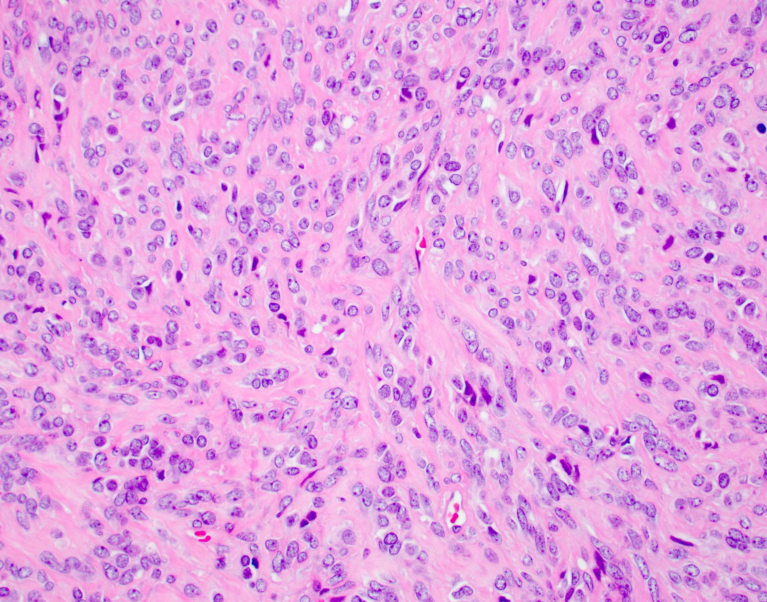
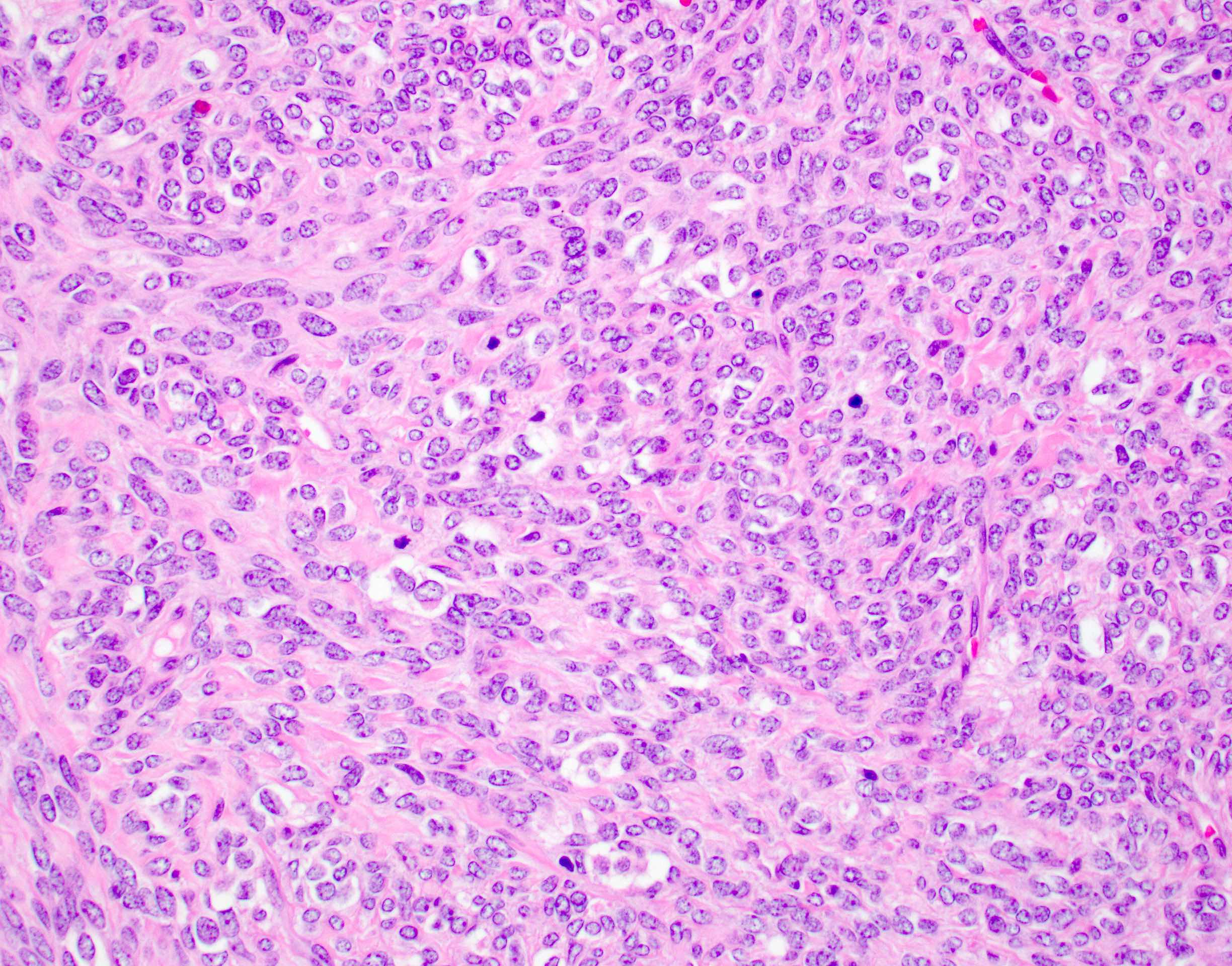
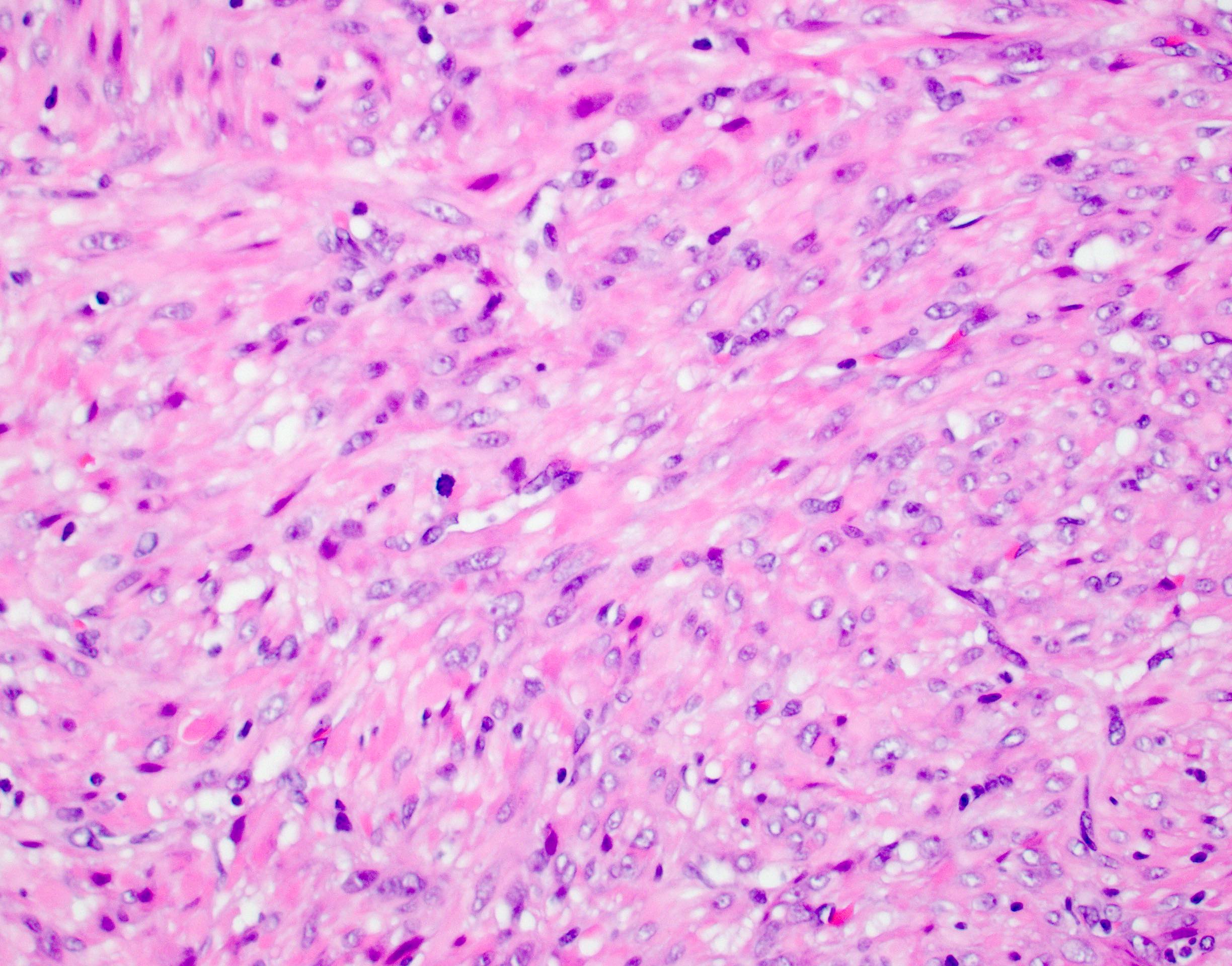
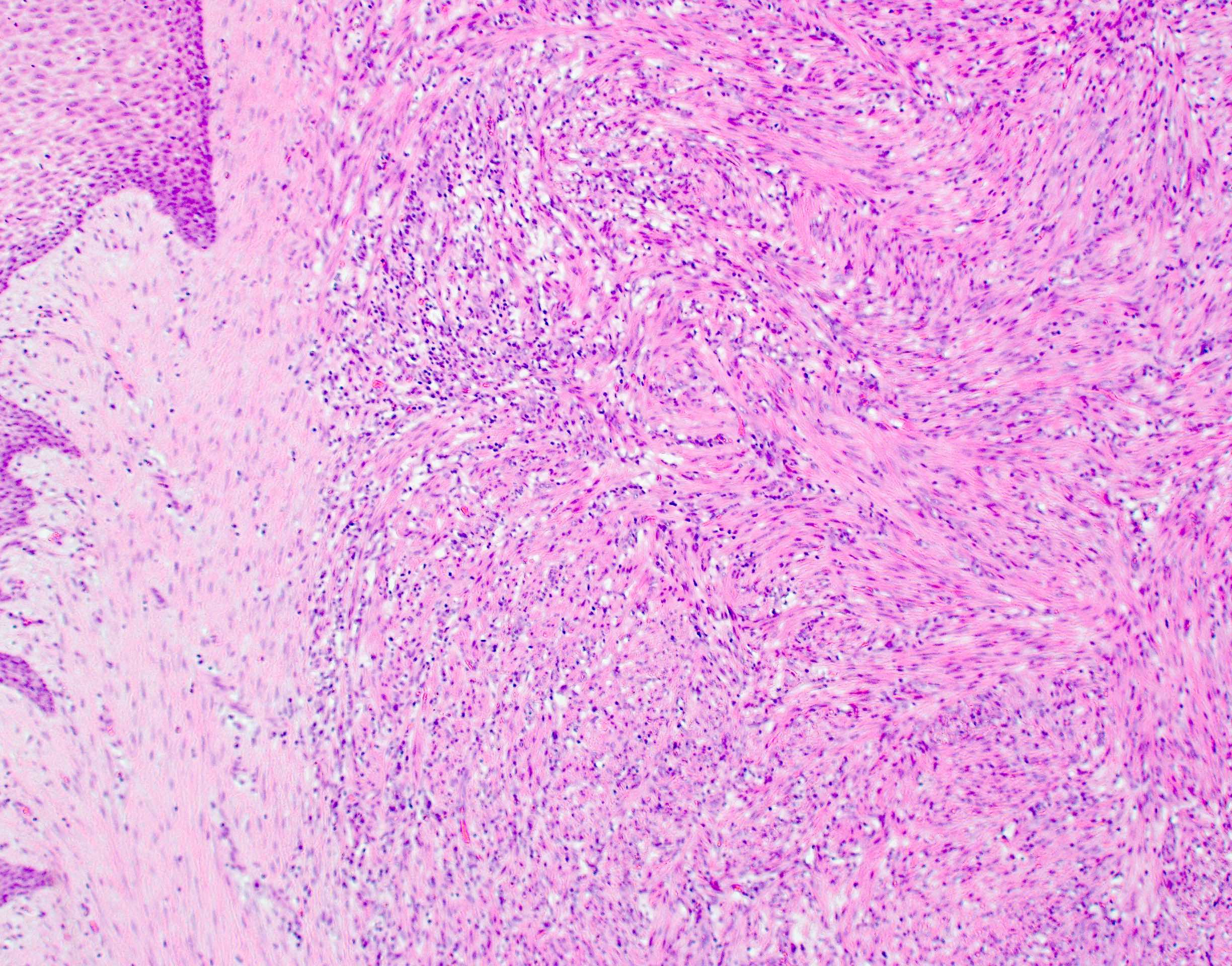
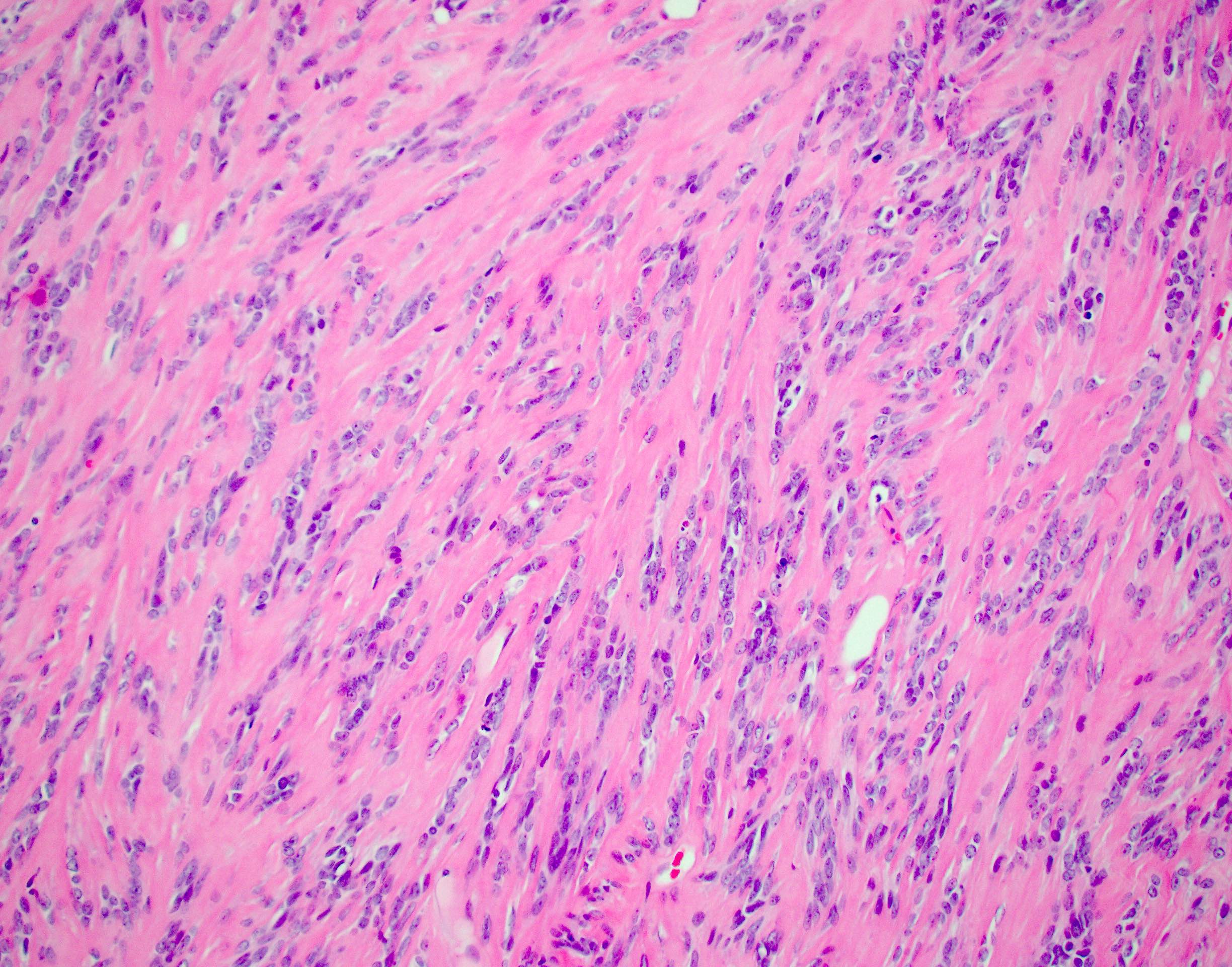
- 均一的卵圆形至梭形细胞排列成短束。
- 病变细胞核形态温和,胞质呈纤维状。
- 无明显细胞异型性。
- 病变细胞呈血管周围排列。
- 模糊的巢状结构,伴丰富血管网。
- 小结节可呈向血管腔内凸出的形态。
- 细胞密度和核分裂率不等。
- 无非典型核分裂象。
- 少部分病例核分裂象超过 10 个 / 2 mm²,并有局灶坏死。
- 可见不同程度的胶原性间质和钙化(Mod Pathol 2025;38:100722)。
- 可有梗死区域,伴营养不良性钙化。
镜下(组织学)图像
细胞学描述
- 散在、形态温和的卵圆形至梭形细胞。
细胞学图像
原文含“形态温和的梭形细胞”和“形态温和的卵圆形细胞”图,贡献者 Laura Warmke, M.D.;本站未收录。
阴性染色
- EMA。
- SOX10。
- MyoD1(Am J Surg Pathol 2020;44:1725)。
- CD34:可局灶阳性(Am J Surg Pathol 2017;41:677)。
- 肌细胞生成素(myogenin)。
- HMB45。
- 广谱细胞角蛋白:部分病例可呈局灶至多灶阳性。
- 延胡索酸水合酶(FH)/ 2SC:FH 表达保留且 2SC 染色阴性,有助于排除 FH 缺陷型平滑肌瘤。
- S100:SRF::ICA1L 肿瘤中曾报道不同程度的表达(Am J Surg Pathol 2020;44:55;Biomolecules 2022;12:1678)。
分子 / 细胞遗传学描述
- 建议确认 SRF 基因重排以明确诊断。
- 最常见的融合伙伴包括 RELA、ICA1L、CITED1 / CITED2 和 NCOA2(Am J Surg Pathol 2017;41:677;Am J Surg Pathol 2020;44:55;Pediatr Dev Pathol 2023;26:65;Mod Pathol 2025;38:100722)。
- 其他伙伴包括 MKL2、C3orf62、ARGFXP1、ARNTL2、NFKBIE、STAT6、NCOA3 和 RGAG1(Am J Surg Pathol 2020;44:1725;Pediatr Blood Cancer 2023;70:e30675;Mod Pathol 2025;38:100722;Virchows Arch 2021;478:597;Hum Pathol 2023;138:41;Semin Diagn Pathol 2026;43:150997)。
分子 / 细胞遗传学图像
原文 SRF::ICA1L 融合和 SRF::RELA 融合图托管于其他服务器;本站未收录。
病理报告示例
- 软组织,前臂肿块,切除:SRF 重排肌样肿瘤(见评注)。
- 评注:切片显示境界清楚的肌样肿瘤,由形态单一的梭形细胞构成,具有中等量淡嗜酸性胞质。可见局灶钙化及玻璃样变胶原沉积区。未见明显细胞异型性或肿瘤性坏死。核分裂象不明显。免疫组化显示病变细胞平滑肌肌动蛋白(SMA)、h-caldesmon 和结蛋白阳性,广谱细胞角蛋白、S100 蛋白、肌细胞生成素、MyoD1 和 CD34 基本阴性。此外,分子检测确认存在 SRF::RELA 基因融合。这些结果支持上述诊断,所有对照切片染色均符合预期。
鉴别诊断
低级别平滑肌肉瘤
- 常发生于成人。
- 细长的雪茄状核,核异型和深染,明显多形性。
- 不伴特定基因融合。
先天性 / 婴儿型梭形细胞横纹肌肉瘤
- 胞质少的梭形细胞形成长束。
- 浸润性边界、核异型和横纹肌母细胞分化。
- 发生于婴儿和幼儿,偏好累及躯干和头颈部。
- 存在涉及 VGLL2 / TEAD1::NCOA2 / CITED2 的基因融合。
- 部分病例有 SRF::NCOA1 和 SRF::FOXO1 融合(Genes Chromosomes Cancer 2013;52:538;Am J Surg Pathol 2020;44:607)。
- 结蛋白、MyoD1 和肌细胞生成素阳性。
- 侵袭性低于经典横纹肌肉瘤。
低级别肌纤维母细胞肉瘤
- 常发生于头颈部。
- 存在核异型;不表达 h-caldesmon。
- 不伴特定基因融合。
肌纤维瘤 / 肌周细胞瘤
- 肌周细胞瘤中,形态温和的梭形细胞围绕众多小血管呈同心圆状生长。
- 肌纤维瘤呈双相形态,包括原始细胞区和肌样梭形细胞结节。
- SMA、h-caldesmon 和肌特异性肌动蛋白阳性。
- 以 PDGFRB 突变为特征。
血管球瘤
- 均一的血管球细胞形成巢状、片状和梁状结构。
- 染色质均匀,形态温和。
- SMA 阳性;IV 型胶原呈显著的细胞周围阳性。
- 可有 NOTCH 融合和 BRAF 突变。
EBV 相关平滑肌肿瘤
- 倾向发生于免疫功能低下患者。
- 常有明显淋巴细胞浸润;内脏部位常见。
- EBER 原位杂交阳性。
延胡索酸水合酶(FH)缺陷型平滑肌瘤
- 可散发,也可发生于遗传性平滑肌瘤病和肾细胞癌综合征背景下。
- 肺泡样水肿和鹿角状血管。
- 卵圆形核,核仁嗜酸性且明显,伴核周晕。
- 可有多核细胞和奇异核细胞。
- FH / 2SC:FH 表达保留且 2SC 染色阴性,有助于排除 FH 缺陷型平滑肌瘤。
孤立性纤维性肿瘤(SFT)
- CD34 和 STAT6 弥漫阳性。
- 伴 NAB2::STAT6 基因融合。
- 缺乏双相结构和肌样细胞形态。
补充参考文献
原文该栏目未列出条目。
练习题 1
SRF 重排肌样肿瘤容易误诊为平滑肌肉瘤或伴肌源性分化的肉瘤。与 SRF 重排肌样肿瘤相比,下列哪项更符合平滑肌肉瘤的特征?
- 局灶浸润性生长
- 细胞密度高
- 核分裂活性
- 核多形性
练习题 1 答案
D. 核多形性。SRF 重排肌样肿瘤不应有明显细胞异型性、核多形性或非典型核分裂象。核多形性更符合平滑肌肉瘤。B 不正确,因为 SRF 重排肌样肿瘤可有不同程度的细胞密度。C 不正确,因为其核分裂活性可有不同程度。A 不正确,因为尽管多数 SRF 重排肌样肿瘤境界相对清楚,但常存在局灶浸润性生长。练习题 2
哪种免疫组化表型最符合 SRF 重排肌样肿瘤?
- CD34 和 STAT6 阳性
- 结蛋白和 MyoD1 阳性
- SMA 和 calponin 阳性
- SMA 和 IV 型胶原阳性
练习题 2 答案
C. SMA 和 calponin 阳性。SRF 重排肌样肿瘤通常表达平滑肌标志物,包括平滑肌肌动蛋白(SMA)和 calponin。D 不正确,因为血管球瘤 SMA 和 IV 型胶原阳性。B 不正确,因为具有骨骼肌分化的肿瘤(包括横纹肌肉瘤)表达结蛋白和 MyoD1。A 不正确,因为尽管 SRF 重排肌样肿瘤可局灶表达 CD34,且一例罕见的 SRF::STAT6 融合病例显示 STAT6 染色,但 CD34 和 STAT6 弥漫染色通常见于孤立性纤维性肿瘤。Definition / general
- Usually benign myoid neoplasm defined by SRF fusion
Essential features
- Monomorphic ovoid to spindle cells with fibrillary cytoplasm
- Demonstrate myoid or immature smooth muscle differentiation
- Expression of 1 or multiple smooth muscle markers
- Detection of SRF gene fusion
Terminology
- Myoid neoplasm of somatic soft tissue
ICD coding
- No unique, standalone code
Epidemiology
- Rare
- Wide age range at presentation
- Most cases occur in pediatric patients (Am J Surg Pathol 2020;44:1725)
- Overall relatively equal gender distribution
Sites
- Anatomic distribution is quite broad
- Most common in deep soft tissue
- May involve subcutis
- Often involves the head and neck, trunk and extremities (Am J Surg Pathol 2017;41:677, Am J Surg Pathol 2020;44:1725, Pediatr Dev Pathol 2023;26:65, Mod Pathol 2025;38:100722)
- Rare cases reported in visceral organs (Am J Surg Pathol 2017;41:677)
Pathophysiology
- SRF gene encodes for serum response factor, a transcription factor that regulates multiple genes involved in cardiac, skeletal and smooth muscle development (Dev Biol 1998;194:18, Eur J Transl Myol 2016;26:6008)
- Most tumors harbor similar SRF breakpoints, resulting in SRF mRNA upregulation at the transcription level
Etiology
- Unknown
Diagnosis
- Myoid or immature smooth muscle differentiation
- Expression of 1 or multiple smooth muscle markers (Semin Diagn Pathol 2026;43:150997)
- Detection of SRF gene fusion
Radiology description
- MRI shows an ovoid mass with hyperintense signal on T2 FS (fat suppressed) and heterogeneous contrast enhancement on T1 FS (Virchows Arch 2021;478:597)
Prognostic factors
- Most cases follow a benign course
- Behavior in children appears to be indolent
- Only 1 patient to date has developed lung metastases, indicating rare potential for malignant behavior (Am J Surg Pathol 2020;44:55)
Case reports
- 3 month old girl with chest wall soft tissue mass (Am J Surg Pathol 2017;41:677)
- 6 year old boy with right buccal mucosa lesion and 12 year old girl with palmar soft tissue mass (Semin Diagn Pathol 2026;43:150997)
- 15 year old boy with left forearm mass (Virchows Arch 2021;478:597)
Treatment
- Surgical resection
Gross description
- Homogeneous tan-white, nodular appearance
- Most are well circumscribed but not encapsulated
- May show infiltrative borders
- Most measure < 5 cm
- Rare cases are > 15 cm (Am J Surg Pathol 2020;44:55, Am J Surg Pathol 2020;44:1725)
Gross images
Gross image of an SRF::ICA1L positive tumor from the original topic was hosted on other servers and was not archived locally.
Microscopic (histologic) description
- Uniform ovoid to spindle cells arranged in short fascicles
- Lesional cells have bland nuclei and fibrillary cytoplasm
- No marked cytologic atypia
- Perivascular arrangement of lesional cells
- Vague nested pattern with a rich vascular network
- Small nodules may appear to bulge into vascular lumina
- Variable cellularity and mitotic rate
- Lack atypical mitotic figures
- Small subset have > 10 mitotic figures per 2 mm 2 and focal necrosis
- Variable collagenous stroma and calcification may be present (Mod Pathol 2025;38:100722)
- May have areas of infarction with dystrophic calcification
Microscopic (histologic) images
Cytology description
- Scattered bland ovoid to spindle cells
Cytology images
Original topic includes “Bland spindle cells” and “Bland ovoid cells” images contributed by Laura Warmke, M.D.; not archived locally.
Negative stains
- EMA
- SOX10
- MyoD1 (Am J Surg Pathol 2020;44:1725)
- CD34 (may be focally positive) (Am J Surg Pathol 2017;41:677)
- Myogenin
- HMB45
- Pancytokeratin: may show focal to multifocal positivity in a subset
- Fumarate hydratase (FH) / 2SC: retained FH expression with negative 2SC staining helps exclude FH deficient leiomyoma
- S100: variable expression has been reported in SRF::ICA1L tumors (Am J Surg Pathol 2020;44:55, Biomolecules 2022;12:1678)
Molecular / cytogenetics description
- Confirmation of SRF gene rearrangement recommended for definitive diagnosis
- Most common gene partners include RELA, ICA1L, CITED1 / CITED2 and NCOA2 (Am J Surg Pathol 2017;41:677, Am J Surg Pathol 2020;44:55, Pediatr Dev Pathol 2023;26:65, Mod Pathol 2025;38:100722)
- Alternative partners include MKL2, C3orf62, ARGFXP1, ARNTL2, NFKBIE, STAT6, NCOA3 and RGAG1 (Am J Surg Pathol 2020;44:1725, Pediatr Blood Cancer 2023;70:e30675, Mod Pathol 2025;38:100722, Virchows Arch 2021;478:597, Hum Pathol 2023;138:41, Semin Diagn Pathol 2026;43:150997)
Molecular / cytogenetics images
SRF::ICA1L and SRF::RELA fusion images from the original topic were hosted on other servers and were not archived locally.
Sample pathology report
- Soft tissue, forearm mass, excision:
- SRF rearranged myoid neoplasm (see comment)
- Comment: Sections show a circumscribed myoid neoplasm composed of monomorphic spindle cells with a moderate amount of pale eosinophilic cytoplasm. Focal calcification and areas with hyalinized collagen deposition are present. No marked cytologic atypia and no tumor type necrosis is identified. Mitotic figures are inconspicuous. Immunohistochemical stains show that the lesional cells are positive for smooth muscle actin (SMA), h-caldesmon and desmin and essentially negative for pancytokeratin, S100 protein, myogenin, MyoD1 and CD34. Additionally molecular testing confirmed the presence of a SRF::RELA gene fusion. These results support the above diagnosis and all control slides stained appropriately.
Differential diagnosis
Low grade leiomyosarcoma
- Often arises in adults
- Elongated cigar shaped nuclei
- Nuclear atypia and hyperchromasia
- Overt pleomorphism
- Not associated with specific gene fusions
Congenital / infantile spindle cell rhabdomyosarcoma
- Long fascicles of spindle cells with scant cytoplasm
- Infiltrative borders, nuclear atypia and rhabdomyoblastic differentiation
- Occurs in infants and young children
- Preferentially involves the trunk and head and neck
- Gene fusions involving VGLL2 / TEAD1::NCOA2 / CITED2
- Subset with SRF::NCOA1 and SRF::FOXO1 fusions (Genes Chromosomes Cancer 2013;52:538, Am J Surg Pathol 2020;44:607)
- Positive for desmin, MyoD1 and myogenin
- Less aggressive than classic rhabdomyosarcoma
Low grade myofibroblastic sarcoma
- Frequently arises in the head and neck
- Nuclear atypia
- Does not express h-caldesmon
- Not associated with specific gene fusions
Myofibroma / myopericytoma
- Myopericytoma shows bland spindled cells growing in a concentric pattern around numerous small blood vessels
- Myofibroma shows a biphasic appearance with primitive cellular zones and nodules of myoid spindled cells
- Positive for SMA, h-caldesmon and muscle specific actin
- Characterized by PDGFRB mutations
Glomus tumor
- Uniform glomus cells forming nests, sheets and trabeculae
- Chromatin is homogeneous and bland
- Positive for SMA
- Collagen IV shows prominent pericellular positivity
- May harbor NOTCH fusions and BRAF mutations
EBV associated smooth muscle tumor
- Tends to arise in immunocompromised patients
- Often has prominent lymphocytic infiltrate
- Visceral location is common
- Positive for EBER ISH
Fumarate hydratase (FH) deficient leiomyoma
- May be sporadic or occur in the setting of hereditary leiomyomatosis and renal cell carcinoma syndrome
- Alveolar edema and staghorn-like vessels
- Ovoid nuclei with prominent eosinophilic nucleoli surrounded by perinuclear halos
- May have multinucleated cells and cells with bizarre nuclei
- Fumarate hydratase (FH) / 2SC: retained FH expression with negative 2SC staining helps exclude FH deficient leiomyoma
Solitary fibrous tumor (SFT)
- Diffusely positive for CD34 and STAT6
- Associated with NAB2::STAT6 gene fusions
- Lack of both biphasic pattern and myoid cytology
Additional references
No entries listed in this section of the original topic.
Practice question #1
SRF rearranged myoid neoplasms may be easily misdiagnosed as leiomyosarcoma or sarcoma with myogenic differentiation. Which of the following features is more characteristic of leiomyosarcoma than SRF rearranged myoid neoplasms?
- Foci of infiltrative growth
- Hypercellularity
- Mitotic activity
- Nuclear pleomorphism
Practice answer #1
D. Nuclear pleomorphism. SRF rearranged myoid neoplasms should not have marked cytologic atypia, nuclear pleomorphism or atypical mitotic figures. The presence of nuclear pleomorphism is more characteristic of leiomyosarcoma than an SRF rearranged myoid neoplasm. Answer B is incorrect because SRF rearranged myoid neoplasms may show a variable degree of cellularity. Answer C is incorrect because SRF rearranged myoid neoplasms may show a variable degree of mitotic activity. Answer A is incorrect because while most SRF rearranged myoid neoplasms are relatively well circumscribed, foci of infiltrative growth are frequently present.Practice question #2
Which immunohistochemical profile fits best with an SRF rearranged myoid neoplasm?
- Positive for CD34 and STAT6
- Positive for desmin and MyoD1
- Positive for SMA and calponin
- Positive for SMA and collagen IV
Practice answer #2
C. Positive for SMA and calponin. SRF rearranged myoid neoplasms are typically positive for smooth muscle markers, including smooth muscle actin (SMA) and calponin. Answer D is incorrect because SMA and collagen IV are positive in glomus tumors. Answer B is incorrect because desmin and MyoD1 are positive in tumors with skeletal muscle differentiation, including rhabdomyosarcoma. Answer A is incorrect because while CD34 may be focally positive in SRF rearranged myoid neoplasms (and a rare case with SRF::STAT6 fusion demonstrated staining with STAT6), diffuse staining with CD34 and STAT6 is typically seen in solitary fibrous tumor.Cite this page: Ghazy A, Warmke L. SRF rearranged myoid neoplasm. PathologyOutlines.com website. https://www.pathologyoutlines.com/topic/softtissuesrfmyoidneoplasm.html. Accessed September 15th, 2026.
English text and images © contributors and PathologyOutlines.com. For personal study only; not medical advice.