- 原文:Case of the Month #566(讨论页;博客 2026-09-18)。
- 贡献:Jeffrey Chang、Farres Obeidin(UCLA);审阅 Nasir Ud Din(University of Miami)。
- 年轻男性运动员股骨髁内病变;诊断为假肌源性血管内皮瘤(PHE),关键分子改变为 FOSB 重排。
感谢 Dr. Jeffrey Chang 与 Dr. Farres Obeidin(University of California, Los Angeles, Los Angeles, California, USA)贡献本病例与讨论,并感谢 Dr. Nasir Ud Din(University of Miami, Florida, USA)审阅讨论。
临床病史
一名 20 岁大学橄榄球运动员,因右膝疼痛 9 个月就诊于家庭医生。患者称疼痛始于一次比赛中扭伤后,保守治疗未能缓解。右膝 MRI 显示股骨外侧髁前方一肿块,T1 低信号、T2 高信号,边界清楚,邻近无骨髓水肿。病灶延伸至前外侧股骨髁关节面,关节面轻度不规则,大小约 1.3 cm × 0.8 cm × 2.1 cm。
影像与镜下图像
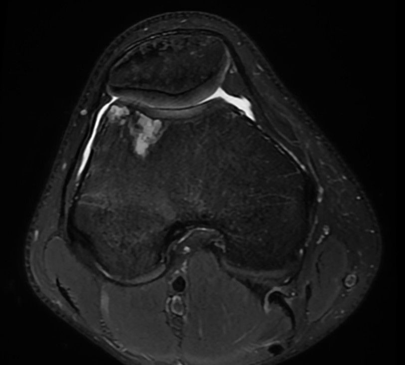
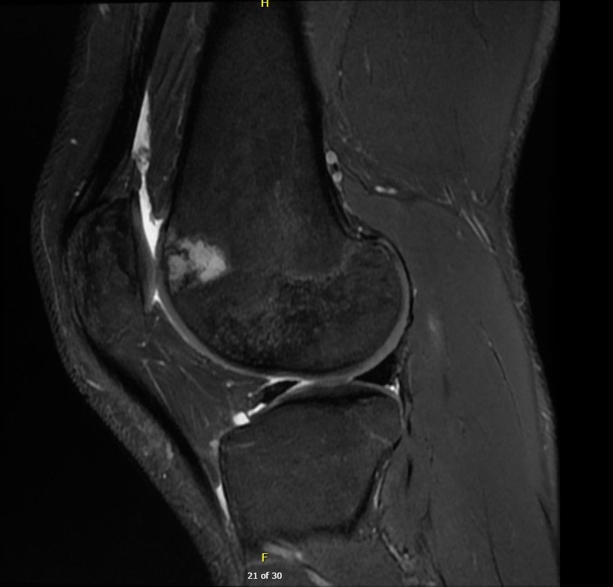
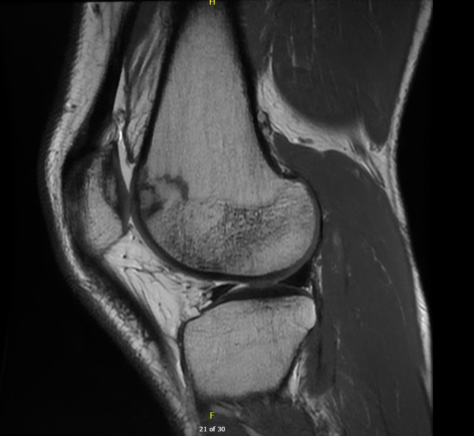
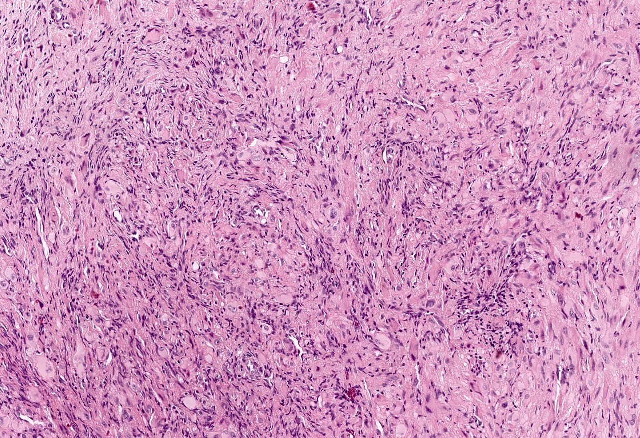
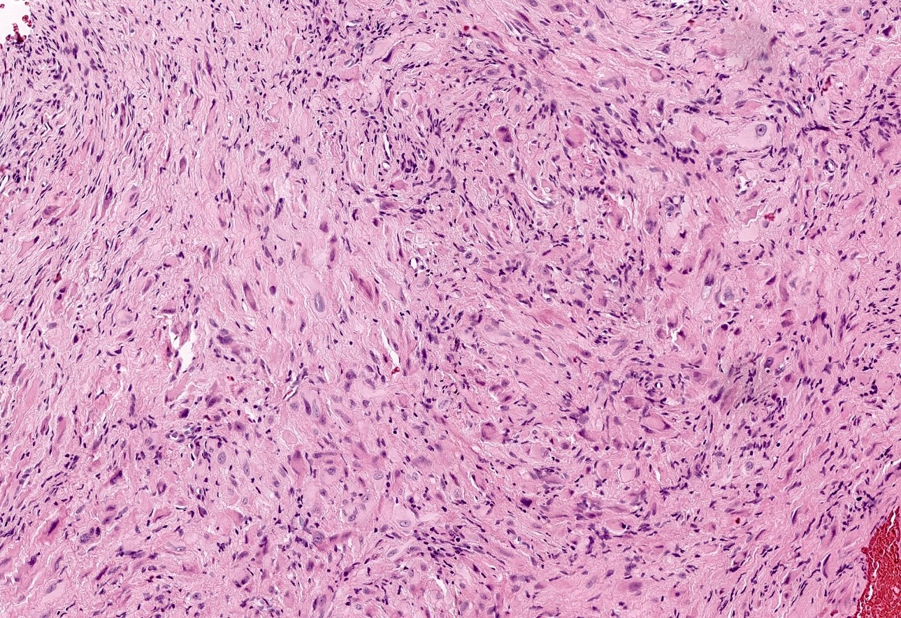
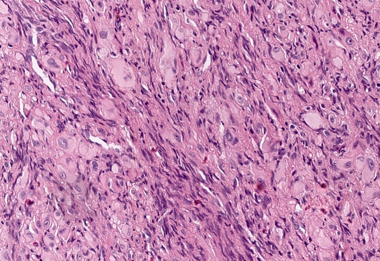
你的诊断是什么?
诊断
假肌源性血管内皮瘤(Pseudomyogenic hemangioendothelioma)
测验题(答案见文末)
与假肌源性血管内皮瘤相关的分子改变是什么?
- ALK 基因重排
- MYC 基因扩增
- FOSB 基因重排
- HEY1::NCOA2 重排
- NTRK1 基因重排
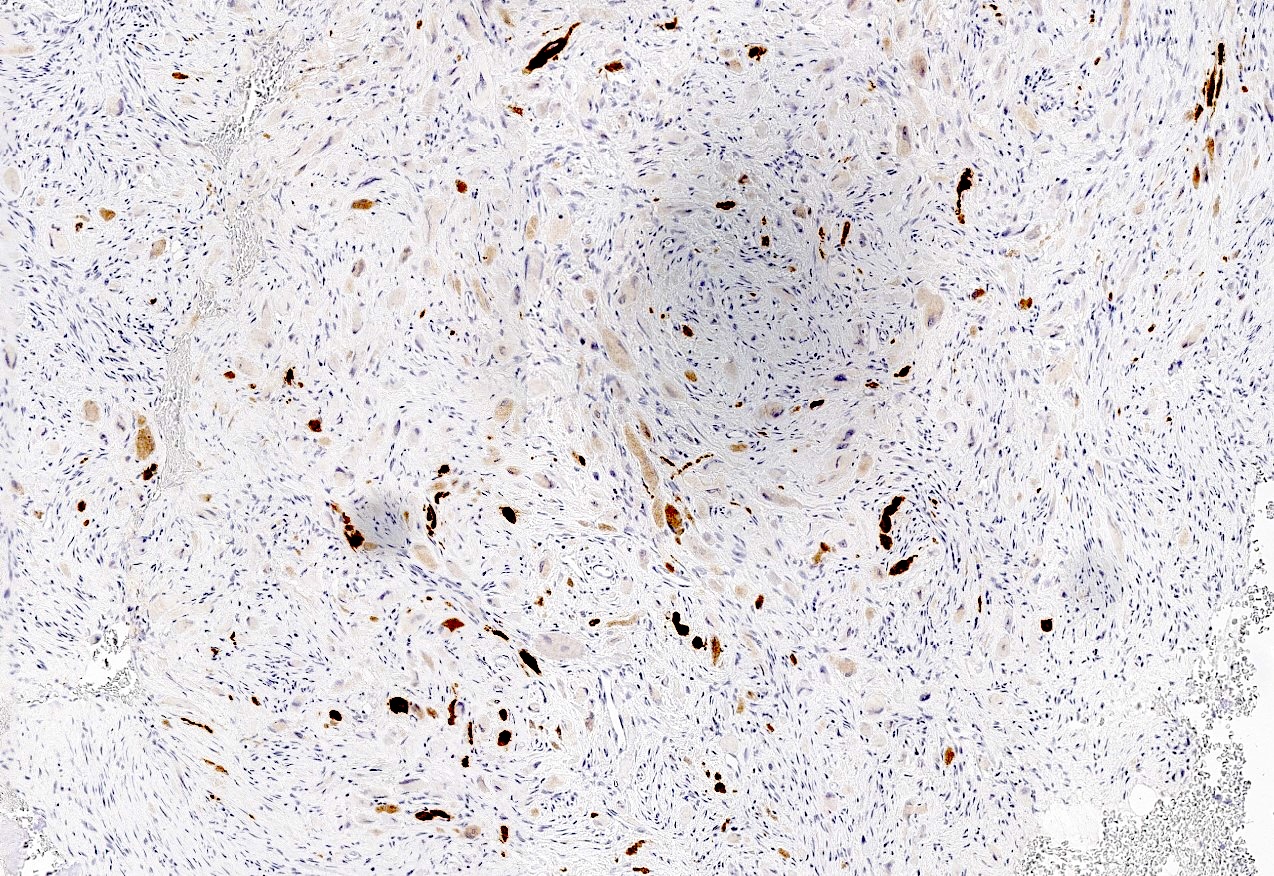
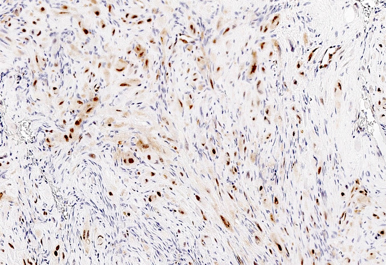
染色
讨论
假肌源性血管内皮瘤(PHE)是一种局部侵袭性血管源性肿瘤,最常见于年轻成年男性(男∶女 ≈ 3∶1)。典型发生于下肢深部软组织。偶可原发于骨内,此时需与原发性骨肿瘤鉴别。原发性骨内 PHE 在骨骼内常呈多灶性,并可出现骨皮质侵蚀。局部复发常见(可达 50%),但全身转移极为罕见。
绝大多数 PHE 存在涉及 FOSB 的基因融合,最常见伴侣基因为 SERPINE1 或 ACTB,导致 FOSB 过表达。FOSB 属 FOS 家族转录因子,调控血管生成组织的细胞增殖与分化。影像上,肿瘤表现为边界清楚的孤立或多灶结节,MRI 上 T1 呈低至等信号,T2 信号强弱不一。
组织学上,肿瘤由肥硕的梭形至上皮样细胞构成,胞质嗜酸性,可类似横纹肌母细胞。通常不见明确成血管结构。免疫组化具特征性:广谱角蛋白(pankeratin)及血管标记 ERG、CD31 一般强阳性(但通常 CD34 阴性);INI1 保留;FOSB 染色可阳性。
治疗以手术切除并争取阴性切缘为主。骨内病变可刮除并长期随访复发。多灶或不可切除病例中,PHE 可对 mTOR 抑制剂、VEGFR1 / PDGFRA 抑制剂以及细胞毒化疗有反应。
测验题答案
C. FOSB 基因重排。假肌源性血管内皮瘤与 FOSB 重排相关,最常见伴侣基因为 SERPINE1 与 ACTB。A 不正确:ALK 融合可见于多种恶性上皮及间叶肿瘤,但未见报道于 PHE。B 不正确:该发现在放疗后血管肉瘤背景下更典型。D 不正确:HEY1::NCOA2 是间叶性软骨肉瘤的典型融合。E 不正确:NTRK 融合常见于一类激酶融合肉瘤(含婴儿型纤维肉瘤、脂肪纤维瘤病样神经肿瘤等)。
- Source: Case of the Month #566 (discussion page; blog 18 September 2026).
- Contributors: Jeffrey Chang and Farres Obeidin (UCLA); reviewed by Nasir Ud Din (University of Miami).
Thanks to Dr. Jeffrey Chang and Dr. Farres Obeidin, University of California, Los Angeles, Los Angeles, California, USA for contributing this case and discussion and Dr. Nasir Ud Din, University of Miami, Florida, USA for reviewing the discussion.
Clinical history
A 20 year old college football player presented to his family physician with a 9 month history of right knee pain. The patient reported noticing the pain after twisting his knee at a football game but the pain hasn't gone away with conservative management. MRI studies of the right knee reveal a mass with a low T1 and high T2 weighted signal within the anterior aspect of the lateral femoral condyle. It appears well delineated without adjacent marrow edema. It extends to the articular surface of the anterolateral femoral condyle, with minimal irregularity, measuring 1.3 cm × 0.8 cm × 2.1 cm.
Radiology and microscopic images
What is your diagnosis?
Diagnosis
Pseudomyogenic hemangioendothelioma
Test question (answer at the end)
What is the molecular alteration associated with pseudomyogenic hemangioendothelioma?
- ALK gene rearrangement
- Amplification of the MYC gene
- FOSB gene rearrangement
- HEY1::NCOA2 rearrangement
- NTRK1 gene rearrangement
Stains
Discussion
Pseudomyogenic hemangioendothelioma (PHE) is a locally aggressive vascular neoplasm that most commonly occurs in young adult males (M:F = 3:1). These tumors typically arise in the deep soft tissue of the lower extremity. However, occasional PHE arise within the bone, raising a differential diagnosis with primary bone tumors. Primary intraosseous PHE is commonly multifocal within the skeleton and can show areas of cortical erosion. Local recurrences are common (up to 50% of cases), though systemic / metastatic disease is very rare. The majority of PHE harbor a gene fusion involving FOSB, most commonly with partner genes SERPINE1 or ACTB, leading to overexpression of FOSB. FOSB is a transcription factor in the FOS family that regulates cellular proliferation and differentiation of angiogenic tissues. Radiographically, these tumors present as well defined, solitary or incontiguous nodules with hypo to isointense T1 signal and variably hyperintense T2 signal on MRI. Histologically, the tumor is composed of plump spindled to epithelioid cells with eosinophilic cytoplasm that may resemble rhabdomyoblasts. Vasoformative features are not typically seen in these lesions. Immunohistochemical stains are characteristic, with generally strong positivity for pankeratins and vascular markers ERG and CD31 (though not CD34). INI1 is retained and a FOSB stain may be positive. PHE is primarily managed with surgical excision with negative margins where possible. Intraosseous tumors may be managed with curettage and long term follow up for recurrences. In multifocal or unresectable disease, PHE has shown response to mTOR inhibitors and VEGFR1 / PDGFRA inhibitors, as well as cytotoxic chemotherapeutic agents.
Test question answer
C. FOSB gene rearrangement. Pseudomyogenic hemangioendothelioma (PHE) is associated with FOSB rearrangements, with SERPINE1 and ACTB as the most common partner genes. Answer A is incorrect because ALK fusions are seen in a broad spectrum of malignant epithelial and mesenchymal neoplasms but have not been seen in PHE. Answer B is incorrect as this finding is typically associated with angiosarcomas in the context of prior radiation therapy. Answer D is incorrect because HEY1::NCOA2 rearrangement is the canonical fusion for mesenchymal chondrosarcoma. Answer E is incorrect as NTRK gene fusions are typical of a subset of kinase fusion sarcomas, a category of molecularly defined tumors that include infantile fibrosarcoma and lipofibromatosis-like neural tumor.