- 27/28 例可判读的子宫颈 NEC 为 HPV 阳性,均见 p16 过表达、Rb 部分缺失。
- 肺小细胞癌对照均为 HPV 阴性,9/10 例 Rb 完全缺失;联合判读 p16 与 Rb 有助于鉴别。
- HPV 阴性子宫颈病例可呈 Rb 保留、p16 局灶及 cyclin D1 阳性;疑难病例需结合 HPV-ISH。
摘要
目的
本研究旨在描述子宫颈神经内分泌癌(UCNEC)中的高危型人乳头瘤病毒(HR-HPV)感染情况,以及 p16、Rb、p53 和 cyclin D1 的表达模式。我们还旨在明确 UCNEC 与肺神经内分泌癌(NEC)之间的异同。
方法与结果
我们对 29 例 UCNEC 进行了组织学复核,采用免疫组织化学(IHC)检测 p16、Rb、p53 和 cyclin D1 的表达模式,并采用 mRNA 原位杂交(ISH)检测 HR-HPV 感染。另纳入 10 例肺小细胞神经内分泌癌(SCNEC)进行比较。组织学上,多数 UCNEC 为合并腺癌或鳞状细胞癌的小细胞神经内分泌癌。28 例 UCNEC 接受了 HR-HPV ISH 检测,其中 27 例(96%)HPV 阳性,1 例(4%)HPV 阴性。全部 HPV 阳性病例(n=27)均表现为 p16 过表达、Rb 部分缺失、p53 野生型模式及 cyclin D1 阴性。唯一的 HPV 阴性病例表现为 p16 局灶阳性、Rb 表达保留、p53 野生型模式及 cyclin D1 阳性。相比之下,全部 10 例肺 SCNEC 均为 HR-HPV 阴性、p16 阳性,Rb 表达完全缺失(n=9)或保留(n=1),并常见 p53 异常表达(n=8;5 例弥漫表达,3 例完全不表达)。
结论
多数 UCNEC 与 HPV 感染相关,其特征为 p16 过表达及 Rb 部分缺失。HPV 阳性 UCNEC 与 HPV 阴性肺 NEC 的 Rb 表达模式不同,提示除 p16 外,Rb 也具有潜在的诊断价值。
缩略语
- ASCL1
- 无刚毛–盾片同源蛋白 1
- FFPE
- 福尔马林固定、石蜡包埋
- FIGO
- 国际妇产科联盟
- HR-HPV
- 高危型人乳头瘤病毒
- IHC
- 免疫组织化学
- LCNEC
- 大细胞神经内分泌癌
- NEUROD1
- 神经源性分化因子 1
- OS
- 总生存期
- POU2F3
- POU 类 2 同源盒蛋白 3
- PPIB
- 肽基脯氨酰异构酶 B
- SCC
- 鳞状细胞癌
- SCNEC
- 小细胞神经内分泌癌
- UCNEC
- 子宫颈神经内分泌癌
- WHO
- 世界卫生组织
- YAP1
- Yes 相关蛋白 1
引言
子宫颈神经内分泌癌(UCNEC)是一种罕见恶性肿瘤,占全部子宫颈癌的 0.5%–5%。[1] UCNEC 生物学行为侵袭性强,容易经淋巴及血行播散,初诊时即有远处转移的情况常见;即使早期确诊,患者死亡率仍然较高。[2–4] 多数 UCNEC 与高危型人乳头瘤病毒(HR-HPV)感染相关,以 16 型和 18 型最常见。[5–7] 根据第 5 版世界卫生组织(WHO)《女性生殖器官肿瘤分类》,UCNEC 在组织学上分为小细胞神经内分泌癌(SCNEC)、大细胞神经内分泌癌(LCNEC),以及混有神经内分泌癌的癌(复合型 NEC)。[8] 复合型 NEC 指非神经内分泌癌与 NEC 成分混合存在。[8]
一般而言,HPV 相关癌表现为 p16 过表达,呈弥漫块状阳性或全细胞染色模式。[9] 我们此前的研究发现,p16 过表达与 Rb 部分(镶嵌状)缺失构成 HPV 相关头颈部鳞状细胞癌(SCC)、[10–12] 子宫颈 SCC[13] 及子宫颈腺癌[14] 的典型免疫组化模式,不过这些肿瘤偶尔也可出现 Rb 完全缺失。在分子层面,HPV 感染细胞中的 HPV E7 蛋白与 Rb 结合,激活 E2F 介导的转录,促进肿瘤发生。[15] 与 HPV E7 结合的 Rb 经泛素–蛋白酶体通路降解。[15] 因而有人提出,Rb 免疫组织化学(IHC)呈部分缺失(镶嵌状染色),可能反映 Rb 降解程度的异质性。[10] 相比之下,HPV 非依赖性癌,包括头颈部 SCC、[10–12] 子宫颈 SCC[13] 和子宫颈腺癌,[14] 通常为 p16 阴性,偶尔为 p16 阳性;无论 p16 表达如何,其 Rb 表达通常保留,完全缺失罕见。目前尚无关于 HPV 相关 NEC 中 Rb 表达模式的详细报道。
NEC 最常发生于肺,通常无 HPV 感染。[6,16] 肺 NEC 经常表现为 Rb 免疫表达完全缺失。[17,18] 最近一项研究报道,多数肺 SCNEC 可归入 Rb 缺陷组,表现为 RB1 突变,常伴 TP53 突变,免疫表型为 Rb 缺失/p16 高表达/cyclin D1 低表达;少数则属于 Rb 功能保留组,表现为 RB1 野生型,常伴 TP53 突变、CDKN2A 突变和 CCDN1 扩增,免疫表型为 Rb 高表达/p16 低表达/cyclin D1 高表达。[18]
本研究旨在阐明 UCNEC 中 HPV 感染的特征及 p16、Rb、p53 和 cyclin D1 的表达模式,并明确子宫颈 NEC 与肺 NEC 之间的异同。
材料与方法
病例选择
回顾性收集 29 例 UCNEC 的福尔马林固定、石蜡包埋(FFPE)活检或手术切除标本,包括 6 例单纯型 SCNEC 和 23 例复合型 NEC(表 1)。全部 29 例患者均于 1999–2024 年在九州大学根据活检或手术标本获得组织病理学诊断。最初从该院病理档案中检索到 31 例 UCNEC。两位重点研究 HPV 相关癌病理的研究者(NY 和 HY)独立复核全部病例。其中 1 例因表达 p40、且不表达神经内分泌标志物而被排除,重新归类为低分化 SCC;另 1 例因标本质量差而被排除,其 Rb IHC 内部阳性对照出现假阴性。最终纳入 29 例 UCNEC。
| 项目 | 全部 (n = 29), n (%) |
|---|---|
| 年龄,中位数(范围),岁 | 46 (28–78) |
| 分期 | |
| I | 15 (52) |
| II | 1 (4) |
| III | 10 (34) |
| IV | 3 (10) |
| 组织学 | |
| 单纯型 SCNEC | 6 (21) |
| 单纯型 LCNEC | 0 (0) |
| 复合型 SCNEC | 22 (76) |
| 复合型 LCNEC | 1 (3) |
| HR-HPV* | |
| 阳性 | 27 (96) |
| 阴性 | 1 (4) |
| 淋巴血管侵犯 | 18 (62) |
| 淋巴结转移 | 8 (28) |
| 远处转移 | 2 (7) |
| 复发 | 13 (45) |
| 死亡 | 10 (34) |
HR-HPV:高危型人乳头瘤病毒;LCNEC:大细胞神经内分泌癌;SCNEC:小细胞神经内分泌癌。* 1 例患者无可用结果。
另检测 10 例肺 SCNEC,作为对照组。全部 10 例患者均于 2015–2023 年在九州大学根据手术标本获得组织病理学诊断。
患者临床资料取自病历。UCNEC 采用 2018 年国际妇产科联盟(FIGO)分期系统进行分期。对于较早的病例(1999–2017 年),按照 2018 年 FIGO 子宫颈腺癌分期系统重新评估分期依据(表 1)。本研究获九州大学机构审查委员会批准,批准号为 22050-01 和 22219。
根据第 5 版 WHO 分类,SCNEC 依据形态学诊断,而 LCNEC 的诊断要求具有相应形态,并表达至少一种神经内分泌标志物(突触素、嗜铬粒蛋白或 CD56)。[8] SCC 成分通过形态学评估及 p40 IHC 表达识别。采用阿利新蓝染色显示胞质内黏液。
免疫组织化学
IHC 使用厚度为 4 μm 的 FFPE 切片,经连续二甲苯及乙醇洗涤完成脱蜡和复水。以 3% H₂O₂ 孵育 10 分钟封闭内源性过氧化物酶,随后进行热诱导抗原修复。按照厂家方案,使用自动染色机(Bond III;Leica Biosystems,澳大利亚墨尔本)进行 IHC,抗体包括:p16(E6H4,预稀释,CIN Histology Kit;Roche,德国海德堡)、Rb(G3-245,稀释度 1/50;BD Pharmingen,美国新泽西州 Franklin Lakes)、突触素(27G12,1/50;Leica Biosystems,英国 Newcastle upon Tyne)、嗜铬粒蛋白 A(LK2H10,1/800;Thermo Fisher Scientific,美国马萨诸塞州 Waltham)、CD56(CD564,1/20;Leica Biosystems,英国 Newcastle upon Tyne)、胰岛素瘤相关蛋白 1(INSM1;A-8,1/3000;Santa Cruz Biotechnology,美国得克萨斯州 Dallas)、p40(BC28,即用型;Nichirei Biosciences,日本东京)、p53(Pab1801,1/2000;Santa Cruz Biotechnology,美国得克萨斯州 Dallas),以及 cyclin D1(SP4,1/75;Abcam,英国剑桥)。
对于突触素、嗜铬粒蛋白 A、CD56、INSM1、p40 和 cyclin D1,按免疫反应阳性肿瘤细胞的比例分为阴性(0%)、局灶阳性(1% 至不足 10%)和阳性(≥10%)。
提示 TP53 突变的 p53 异常表达定义为过表达(超过 80% 的肿瘤细胞核呈强阳性染色),或完全不表达模式(内部对照保留,但肿瘤细胞无核染色,或出现明确的胞质染色)。其余病例均归为 p53 野生型模式。[19]
p16 阳性定义为几乎全部肿瘤细胞出现强而弥漫的细胞核及胞质染色,即“块状”阳性。[20]
采用既往报道的评分体系,将 Rb 核表达分为完全缺失(阳性比例 <10%)、表达保留(>90%)及部分缺失(10%–90%)。[11,14] 我们既往研究显示,对 Rb 表达模式的评估具有较高的观察者间重复性。[11]
高危型 HPV mRNA 原位杂交
按照厂家方案,采用针对 18 种 HR-HPV 型别(16、18、26、31、33、35、39、45、51、52、53、56、58、59、66、68、73 和 82 型)的 E6/E7 mRNA 探针组(Advanced Cell Diagnostics,美国加利福尼亚州 Newark)及 RNAscope 2.5HD 检测试剂盒(Advanced Cell Diagnostics),通过原位杂交(ISH)检测 HPV RNA。只要出现任何核内或胞质点状信号,即判为阳性。[21] 口咽 SCC 标本用作 HPV-ISH 外部阳性对照。[10] 同时验证肽基脯氨酰异构酶 B(PPIB)mRNA 的表达,作为内部对照。
统计学分析
采用单因素及多因素 Cox 比例风险回归分析识别预后因素。采用 Kaplan–Meier 分析和 log-rank 检验估计并比较生存曲线。总生存期(OS)定义为从治疗首日至死亡的时间;对于删失病例,则计算至末次随访日期。统计分析使用 JMP19(SAS Institute,美国北卡罗来纳州 Cary)完成。P <0.05 视为差异具有统计学意义。
结果
UCNEC 的临床病理学特征
表 1 汇总了 29 例 UCNEC 的临床病理学特征。患者年龄为 28–78 岁,中位年龄 46 岁;52% 为 I 期。18 例(62%)存在淋巴血管侵犯,8 例(28%)存在淋巴结转移,2 例(7%)存在远处转移。OS 为 1–241 个月,中位 48 个月。
UCNEC 中的 HPV 感染
28 例成功完成 HR-HPV ISH 检测,其中 27 例(96%)HPV 阳性,1 例(4%)HPV 阴性(表 1)。余下 1 例未检出 HPV 和内源性 PPIB mRNA 信号,因此结果无法判读。
UCNEC 的组织学亚型
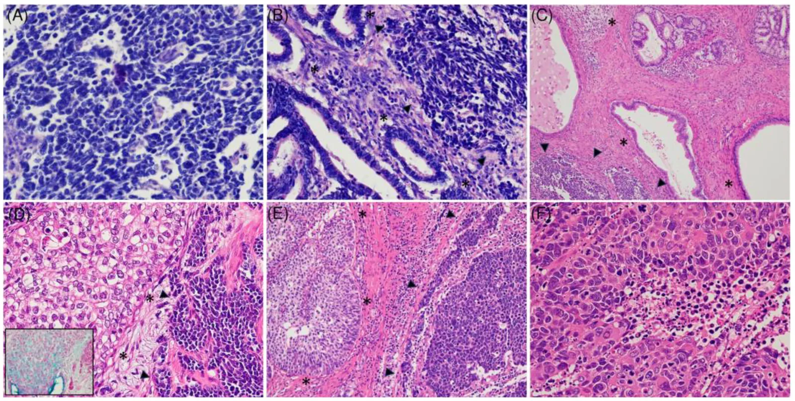
UCNEC 在组织学上分为单纯型 SCNEC(n=6)和复合型 NEC(n=23);后者包括复合型 SCNEC(n=22)和复合型 LCNEC(n=1)(图 1、表 1)。6 例单纯型 SCNEC 中,1 例根据活检标本诊断,其余 5 例根据手术标本诊断。
图 2 汇总了组织学亚型及 HPV 感染状态。HPV 阳性病例(n=27)包括单纯型 SCNEC(n=4)及复合型 NEC(n=23)。HPV 阴性病例(n=1)为单纯型 SCNEC,HPV 状态不确定的病例(n=1)也为单纯型 SCNEC。
22 例复合型 SCNEC 中,全部非 NEC 成分均为 HPV 相关癌,包括腺癌(n=11)、SCC(n=9)、腺鳞癌(n=1)及腺样囊性癌样癌(n=1)。腺癌进一步分为普通型(n=7)、浸润性分层产黏液癌(iSMILE;n=2)、黏液性非特指型(NOS;n=1)及原位腺癌(AIS;n=1)。11 例 IHC 显示 p40 部分或弥漫阳性,结合形态学分别归类为 SCC(n=9)、腺鳞癌(n=1)和腺样囊性癌样癌(n=1)。对 2 例形态提示 iSMILE 的病例进行了阿利新蓝染色,以证实细胞内黏液生成。1 例复合型 LCNEC 含 HPV 相关普通型腺癌。总体而言,复合型 NEC 的腺癌成分以普通型腺癌最常见(8/12 例)。
UCNEC 中的神经内分泌及细胞周期标志物
UCNEC 中,肿瘤表达突触素(19/29 例;66%)、嗜铬粒蛋白 A(6/29 例;21%)、CD56(19/29 例;66%)及 INSM1(21/29 例;72%)(图 2)。复合型 NEC 中,当非 NEC 癌成分为 SCC 时,神经内分泌标志物(突触素、嗜铬粒蛋白 A、CD56 和 INSM1)的表达倾向于较低(图 2)。
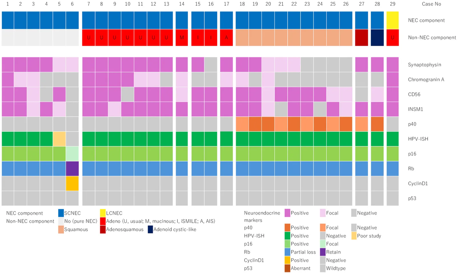
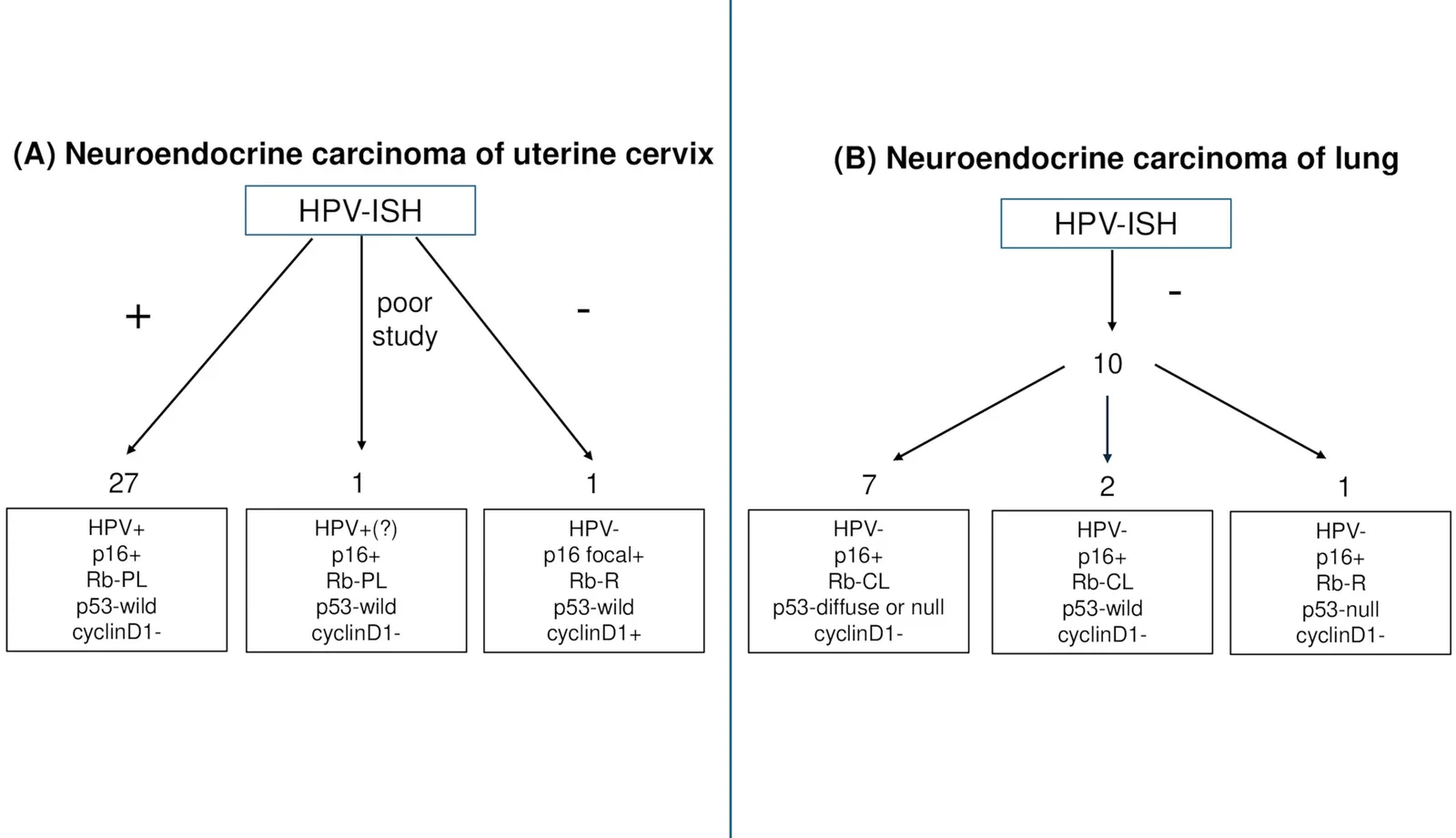
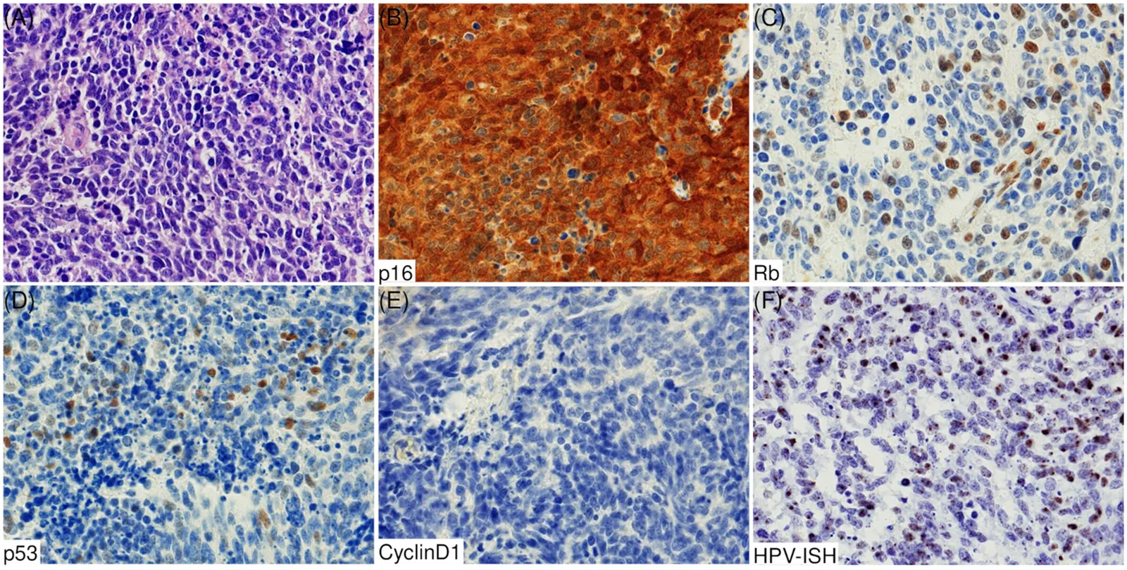
细胞周期标志物的结果汇总于图 3。HPV 阳性病例(n=27),包括单纯型及复合型 NEC,均表现为 p16 弥漫性核与胞质过表达(“块状”阳性)、Rb 部分缺失、p53 野生型模式及 cyclin D1 阴性(图 4、5)。这些标志物在 NEC 与非 NEC 成分中的表达模式基本一致(图 5)。26 例 HPV 阳性病例的 NEC 成分显示常规 SCNEC 形态;余下 1 例为复合型 LCNEC(病例 29),其免疫组化标志物表达模式与 HPV 相关 SCNEC 基本相同(图 2、3)。
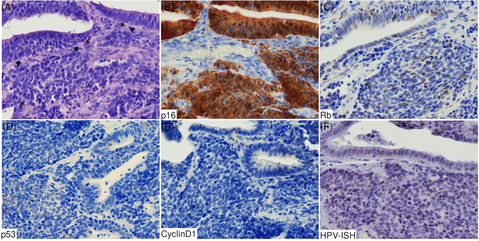
HPV 阴性病例(病例 6)表现为 p16 斑片状表达、Rb 表达保留、p53 野生型模式及 cyclin D1 阳性(图 2、6)。该病例的形态与 HPV 相关 SCNEC 相同(图 6)。
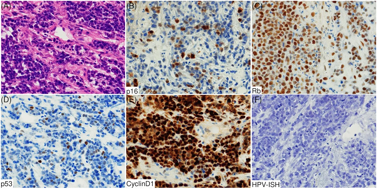
在无法获得 HPV-ISH 结果的病例(病例 5)中,p16、Rb、p53 和 cyclin D1 的表达模式与 HPV 阳性病例相似,提示该肿瘤很可能是 HPV 相关 SCNEC(图 2、3)。
在部分缺失(PL)模式的病例中,Rb 表达指数为 20%–60%,中位数为 30%(表 S1)。Rb 表达保留的病例中,几乎全部肿瘤细胞均表达 Rb,比例可达 100%;Rb 完全缺失的病例中,几乎全部肿瘤细胞均明确不表达 Rb,阳性比例接近 0%。
肺 SCNEC 的组织学亚型及 HPV 感染
全部肺 NEC 病例(n=10)在组织学上均归类为单纯型 SCNEC。无一例显示 HPV 感染证据。
肺 SCNEC 中的免疫组化标志物
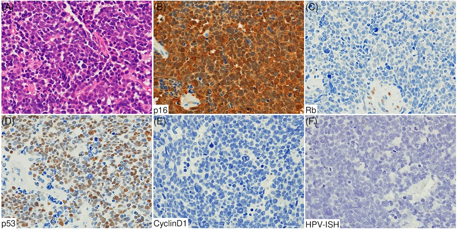
免疫组化分析显示,10/10 例(100%)p16 过表达,9/10 例(90%)Rb 完全缺失,1/10 例(10%)Rb 表达保留,10/10 例(100%)cyclin D1 阴性,8/10 例(80%)p53 异常表达(弥漫或完全不表达模式);余下 2/10 例(20%)为 p53 野生型模式(图 7)。
根据 Rb 和 p53 表达差异,将 10 例肺 SCNEC 的免疫表型分为三种模式:7 例(70%)Rb 完全缺失并伴 p53 异常模式(5 例弥漫,2 例完全不表达);2 例(20%)Rb 完全缺失并伴 p53 野生型模式;余下 1 例(10%)Rb 表达保留并伴 p53 异常模式(完全不表达)(图 3)。
UCNEC 的临床病理因素与预后关联
单因素 Cox 比例风险分析显示,年龄较大(≥50 岁,P=0.0401)和分期较晚(III+IV 期,P=0.0366)与 UCNEC 患者较短的 OS 显著相关(表 S2)。
Kaplan–Meier 生存分析显示,子宫颈单纯型 SCNEC 与复合型 NEC 的 OS 无差异(P=0.5960)。同样,含腺癌成分与含 SCC 成分的子宫颈复合型 NEC 之间,OS 也无差异(P=0.4568)。
讨论
在本组具有 18 种 HR-HPV mRNA ISH 可用结果的 UCNEC 患者中,绝大多数病例(27/28,96%)为 HR-HPV 阳性。既往报道的 UCNEC HR-HPV 感染率为 65.6%–93.8%。[22–25] 这种差异的一个可能原因是检测方法与样本不同:既往研究分别采用全外显子组测序(新鲜冷冻及 FFPE 组织)、[22] 针对 HPV 16 和 18 型的 HPV mRNA ISH(FFPE 组织)、[23] 检测 28 种 HPV 基因型的 HPV 分型聚合酶链反应(PCR;FFPE 组织)、[24] 基于 PCR 检测 14 种高危及 18 种低危 HPV 基因型(液基细胞学标本),[25] 或针对 100 种 HPV 基因型的新一代测序(FFPE 组织)。[25] 其他可能解释还包括病例选择偏倚(我们仅分析了 29 例)及地域差异。
本组病例中,无论组织学亚型如何,HR-HPV 感染均十分常见。普通型腺癌是复合型 UCNEC 最常见的非 NEC 成分。[8] 虽然本组也常见普通型腺癌,但我们还识别出其他 HPV 相关腺癌亚型,包括黏液性 NOS 型(n=1)和 iSMILE(n=2)。[23,26,27] 据我们所知,此前尚无 NEC 与黏液性 NOS 型腺癌复合的报道。在一项大规模 iSMILE 病例系列中,仅有 1 例 NEC 与 iSMILE 复合的记录。[28] 与既往报道不同,[8] 本组复合型 UCNEC 的非 NEC 成分中,41% 为 SCC。
本研究中,全部 HPV 阳性 UCNEC 的 NEC 及非 NEC 成分均显示 p16 过表达和 Rb 部分缺失。虽然仅发现 1 例 HPV 阴性 UCNEC(单纯型 SCNEC),但其表现为 p16 局灶表达(非“块状”阳性)及 Rb 表达保留。本研究观察到的 HPV 感染与 p16/Rb 表达之间的关系,与我们此前在子宫颈 SCC、[13] 子宫颈腺癌[14] 及头颈部 SCC[10–12] 中的发现一致。在子宫颈 SCC 细胞系中,HPV E7 蛋白与 Rb 蛋白结合,并通过泛素–蛋白酶体通路诱导 Rb 蛋白降解。[15] 因此,我们推测 Rb 降解的异质性可能解释 HPV 相关 SCC 和腺癌中 Rb IHC 的镶嵌状表达(部分缺失)模式。[10–14] 同样,HPV 相关 UCNEC 也可能通过类似机制呈现 Rb 部分缺失,但这一假说仍需进一步实验研究验证。另一方面,RB1 基因突变在 UCNEC 中罕见,仅见于 7%–10% 的病例。[29–31] Ordulu 等对 14 例 UCNEC 的分析发现,4 例存在 RB1 异常,其中包括全部 3 例 HPV 阴性病例(100%),而 11 例 HPV 阳性病例中仅 1 例(9%)存在异常。[23] 这些发现提示,RB1 异常不太可能解释 HPV 阳性 UCNEC 中 Rb 表达的降低。
关于其他器官 HPV 相关 NEC 的 Rb 表达模式,详细分析仍然有限。一项结直肠 NEC 研究将 HPV 相关癌描述为“Rb 阳性”,[32] 但其发表的图像似乎显示 Rb 部分缺失。因此,HPV 相关 NEC 可能无论发生于何种器官,都表现为 p16 过表达及 Rb 部分缺失。
本研究未在任何肺 SCNEC 中检出 HR-HPV 感染,与全球既往研究的结果(0%–33%)一致。[6,16,33] 这些发现证实,肺 SCNEC 通常不存在 HR-HPV 感染。
RB1 和 TP53 异常在肺 SCNEC 的发生中发挥关键作用。近年来,已有关于这些基因变异的报道;多数肺 SCNEC 属于 Rb 缺陷组,表现为 RB1 突变,常伴 TP53 共突变,免疫表型为 Rb 缺失/p16 高表达/cyclin D1 低表达。[18] 在肺 SCNEC 中,RB1 改变(突变和/或缺失)导致 IHC 上 Rb 完全缺失及 p16 过表达。类似地,本组 10 例肺 SCNEC 中有 9 例(90%)表现为 p16 过表达、Rb 完全缺失及 cyclin D1 阴性的免疫组化模式,并常伴 p53 异常模式。另一方面,少数肺 SCNEC 属于 Rb 功能保留组,表现为 RB1 野生型,常伴 TP53 突变、CDKN2A 突变、CCND1 扩增,免疫表型为 Rb 高表达/p16 低表达/cyclin D1 高表达。[18] 本组肺 SCNEC 中有 1 例 Rb 表达保留、p16 过表达且 cyclin D1 阴性。肺 SCNEC 中偶尔可观察到突变但无功能的 Rb 蛋白表达。[18] 虽然我们未分析 RB1 突变,但从分子角度看,这一病例可能应视为功能性 Rb 缺陷。值得注意的是,本组 29 例 UCNEC 中有 1 例(3%)为 HPV 阴性,表现为 Rb 表达保留(高)、p16 局灶表达(低)及 cyclin D1 阳性(高)。这一免疫组化模式符合肺 SCNEC 中所描述的 Rb 功能保留组特征。这提示,子宫颈与肺的少数 NEC 似乎属于 HPV 阴性/Rb 功能保留的分子亚型。然而,这仍属推测,需要对更多 HPV 非依赖性 UCNEC 进行分子遗传学分析。
在遗传学层面,肺 SCNEC 几乎普遍存在体细胞 TP53 突变,[34] 而子宫颈 SCNEC 的 TP53 突变较少见(13%–26%)。[29,31] Hillman 等报道,HPV 阳性 UCNEC 中 1/8 例(12.5%)、HPV 阴性 UCNEC 中 1/7 例(14.3%)存在 TP53 突变。[31] 本组 HPV 阳性 UCNEC 未见 p53 异常表达;相反,HPV 阴性肺 SCNEC 中经常出现 p53 异常表达,与既往报道一致。[34]
据报道,HPV 病毒癌蛋白 E6 可使 p53 失活。[35] 因此,在 HPV 相关 UCNEC 中,p53 可能是受到 HPV 癌蛋白 E6 的抑制,而非因 TP53 突变失活,从而促进肿瘤发生。考虑到 TP53 和 RB1 改变在肺 SCNEC 发病机制中的关键作用,UCNEC 中的 HPV 感染可能提供另一种机制:分别通过病毒编码的癌蛋白 E6 和 E7,使 p53 和 Rb 失活。[36] 换言之,无论 HPV 状态如何,Rb–E2F 通路异常及 p53 功能障碍可能在多数 NEC 的发生中发挥核心作用。
本研究的局限是所检测的 UCNEC 与肺 SCNEC 数量相对较少,且未对 RB1、TP53 及其他细胞周期调控因子进行遗传学分析。应谨慎解释本研究结果,并需要更大队列的进一步分析,以阐明 HPV 相关及 HPV 非依赖性癌的肿瘤发生分子机制和不同的 Rb 表达模式。
作为诊断筛查工具,p16 阳性/Rb 部分缺失模式可能有助于提示 NEC 中的 HPV 感染。相比之下,如前所述,HPV 阴性肺 SCNEC 表现为 p16 过表达和 Rb 完全缺失。因此,联合评估 p16 与 Rb 表达谱可能有助于鉴别诊断,尤其是判断可能的原发部位。不过,一个诊断陷阱是部分 HPV 阳性头颈部 SCC 也可表现为 p16 过表达和 Rb 完全缺失,这可能增加转移性病变的鉴别难度。对于此类疑难病例,应结合 p40、神经内分泌标志物 IHC 及 HPV-ISH 确认诊断。
肺 SCNEC 的分子分型正受到广泛关注。多项研究发现,按新型神经内分泌标志物表达划分的亚组之间存在预后差异,这些标志物包括神经源性分化因子 1(NEUROD1)、无刚毛–盾片同源蛋白 1(ASCL1)、POU 类 2 同源盒蛋白 3(POU2F3)及 Yes 相关蛋白 1(YAP1)。[37–40] 值得注意的是,NEUROD1 亚组占 UCNEC 病例的 50%,且与不良预后相关。[24] 仍需进一步研究明确 UCNEC 分子分型的临床及治疗意义。
总之,本研究描述了 UCNEC 的 HPV 感染状态及 p16、Rb、p53 和 cyclin D1 的表达模式,并界定了 UCNEC 与肺 NEC 之间的差异。日本患者中,多数 UCNEC 的特征为 HPV 感染伴 p16 过表达、Rb 部分缺失及 cyclin D1 阴性,这可能源于 HPV 病毒癌蛋白 E7 介导的 Rb 降解。少数 UCNEC 可能对应肺 SCNEC 的 Rb 功能保留亚型,其特征为 HPV 阴性/Rb 表达保留/p16 低表达/cyclin D1 高表达。
- 27/28 evaluable cervical NECs were HPV-positive, with p16 overexpression and partial Rb loss.
- All pulmonary small cell carcinoma controls were HPV-negative; 9/10 showed complete Rb loss, supporting combined p16/Rb assessment.
- The HPV-negative cervical case retained Rb, with focal p16 and positive cyclin D1; equivocal cases require HPV-ISH correlation.
Abstract
Aims
This study aimed to characterize high-risk human papillomavirus (HR-HPV) infection and the expression patterns of p16, Rb, p53 and cyclin D1 in uterine cervical neuroendocrine carcinomas (UCNECs). We also aimed to clarify the similarities and differences between UCNECs and pulmonary neuroendocrine carcinomas (NECs).
Methods and results
We examined the expression patterns of p16, Rb, p53, and cyclin D1 by immunohistochemistry (IHC) and HR-HPV infection by mRNA in situ hybridization (ISH) in 29 UCNEC cases, along with histological review. Ten pulmonary small cell neuroendocrine carcinomas (SCNECs) were included for comparison. Histologically, most UCNECs were SCNECs combined with adenocarcinoma or squamous cell carcinoma. HR-HPV ISH was performed in 28 UCNEC cases; 27 (96%) were HPV-positive and 1 (4%) was HPV-negative. All HPV-positive cases ( n = 27) showed p16 overexpression, partial Rb loss, a wildtype p53 pattern, and negative cyclin D1 expression. The single HPV-negative case showed focal p16 positivity, retained Rb expression, a wildtype p53 pattern and positive cyclin D1 expression. In contrast, all 10 pulmonary SCNEC cases were negative for HR-HPV and positive for p16, showing complete loss ( n = 9) or retained ( n = 1) Rb expression, and often aberrant p53 expression ( n = 8; 5 diffuse and 3 null).
Conclusions
Most UCNECs are associated with HPV infection and characterized by p16 overexpression as well as partial loss of Rb. The Rb expression pattern differs between HPV-positive UCNECs and HPV-negative pulmonary NECs, suggesting the potential diagnostic utility of Rb as well as p16.
Abbreviations
- ASCL1
- achaete-scute homologue 1
- FFPE
- Formalin-fixed, paraffin-embedded
- FIGO
- International Federation of Gynecology and Obstetrics
- HR-HPV
- high-risk human papillomavirus
- IHC
- immunohistochemistry
- LCNEC
- large cell neuroendocrine carcinoma
- NEUROD1
- neurogenic differentiation factor 1
- OS
- Overall survival
- POU2F3
- POU class 2 homeobox 3
- PPIB
- Peptidylprolyl isomerase B
- SCC
- squamous cell carcinoma
- SCNEC
- small cell neuroendocrine carcinoma
- UCNEC
- uterine cervical neuroendocrine carcinoma
- WHO
- World Health Organization
- YAP1
- yes-associated protein 1
Introduction
Neuroendocrine carcinoma of the uterine cervix (UCNEC) is a rare malignancy, accounting for 0.5%–5% of all uterine cervical cancers. 1 UCNEC is biologically aggressive, with a high propensity for lymphatic and haematogenous spread. Distant metastasis at presentation is common. Mortality is high, even among patients diagnosed at an early stage. 2 - 4 Most UCNECs are associated with high-risk human papillomavirus (HR-HPV) infection, most commonly types 16 and 18. 5 - 7 According to the fifth edition of the World Health Organization (WHO) Classification of Female Genital Tumours, UCNECs are histologically subclassified into small cell neuroendocrine carcinoma (SCNEC), large cell neuroendocrine carcinoma (LCNEC) and carcinoma admixed with neuroendocrine carcinoma (combined NEC). 8 Combined NEC refers to a non-neuroendocrine carcinoma admixed with a NEC component. 8
In general, HPV-associated carcinomas exhibit p16 overexpression with a diffuse block positivity or pan-cellular staining pattern. 9 Our previous studies revealed that p16 overexpression and partial (mosaic) loss of Rb constitute a typical immunohistochemical pattern in HPV-associated head and neck squamous cell carcinoma (SCC), 10 - 12 uterine cervical SCC 13 and uterine cervical adenocarcinoma, 14 although complete loss of Rb occasionally occurs in these tumours. At the molecular level, in HPV-infected cells, the HPV E7 protein binds to Rb, activates E2F-mediated transcription and contributes to tumorigenesis. 15 HPV E7-bound Rb is degraded through the ubiquitin–proteasome pathway. 15 Thus, it is hypothesized that the partial loss (mosaic staining) pattern of Rb immunohistochemistry (IHC) may represent heterogeneous levels of Rb degradation. 10 In contrast, HPV-independent carcinomas, including head and neck SCC, 10 - 12 uterine cervical SCC, 13 and uterine cervical adenocarcinoma, 14 are usually p16-negative but occasionally p16-positive, and show retained Rb expression (with rare cases of complete loss) regardless of p16 expression. There have been no detailed reports on Rb expression patterns in HPV-associated NEC.
NECs most often occur in the lung and are usually negative for HPV infection. 6 , 16 Pulmonary NECs frequently exhibit complete loss of Rb immunoexpression. 17 , 18 A recent study reported that most pulmonary SCNECs can be subclassified into an Rb-deficient group ( RB1 mutation often with TP53 mutation, and an Rb-loss/p16-high/cyclin D1–low immunophenotype), while a small proportion belongs to an Rb-proficient group ( RB1 wildtype, often with TP53 mutation, CDKN2A mutation and CCDN1 amplification, with an Rb-high/p16-low/cyclin D1–high immunophenotype). 18
This study aims to elucidate the characteristics of HPV infection and the expression patterns of p16, Rb, p53 and cyclin D1 in UCNECs. It also seeks to clarify the similarities and differences between uterine cervical NECs and pulmonary NECs.
Materials and methods
Case Selection
Formalin-fixed, paraffin-embedded (FFPE) biopsy or surgically resected specimens were retrospectively collected from 29 UCNECs including 6 pure SCNECs and 23 combined NECs (Table 1 ). All 29 UCNEC patients were histopathologically diagnosed based on biopsy or surgical specimens at Kyushu University between 1999 and 2024. Initially, 31 cases of UCNEC were identified in the hospital's pathology record. All cases were independently reviewed by two investigators (NY and HY) who have a special interest in HPV-associated cancer pathology. One case was excluded because of the presence of p40 expression and absence of neuroendocrine marker expression, and it was reclassified as poorly differentiated SCC. Another case was excluded because of poor sample quality (false negativity for Rb IHC was seen in the internal positive control). Finally, 29 cases of UCNEC were enrolled.
| Factors | All (n = 29), n (%) |
|---|---|
| Age, median years (range) | 46 (28–78) |
| Stage | |
| I | 15 (52) |
| II | 1 (4) |
| III | 10 (34) |
| IV | 3 (10) |
| Histology | |
| Pure SCNEC | 6 (21) |
| Pure LCNEC | 0 (0) |
| Combined SCNEC | 22 (76) |
| Combined LCNEC | 1 (3) |
| HR-HPV* | |
| Positive | 27 (96) |
| Negative | 1 (4) |
| Lymphovascular invasion | 18 (62) |
| Lymph node metastasis | 8 (28) |
| Distant metastasis | 2 (7) |
| Recurrence | 13 (45) |
| Death | 10 (34) |
HR-HPV, high-risk human papillomavirus; LCNEC, large cell neuroendocrine carcinoma; SCNEC, small cell neuroendocrine carcinoma. * Not available for 1 patient.
Ten cases of pulmonary SCNEC were also examined as a control group. All 10 pulmonary SCNEC patients were histopathologically diagnosed based on surgical specimens at Kyushu University between 2015 and 2023.
Patients' clinical data were obtained from their medical records. UCNECs were staged using the 2018 International Federation of Gynecology and Obstetrics (FIGO) staging system. For older cases (1999–2017), staging evidence was re-evaluated according to the 2018 FIGO staging system for uterine cervical adenocarcinomas (Table 1 ). This study was approved by the Institutional Review Board of Kyushu University (approval nos. 22050-01 and 22219).
According to the fifth edition of the WHO classification, SCNECs were diagnosed based on morphology, whereas LCNECs required morphology and expression of at least one neuroendocrine marker (synaptophysin, chromogranin or CD56). 8 The SCC component was identified by morphological evaluation and p40 IHC expression. Intracytoplasmic mucin was demonstrated by Alcian blue staining.
Immunohistochemistry
IHC was performed on 4-μm-thick FFPE sections, which were deparaffinized and rehydrated through serial xylene and ethanol washes. Endogenous peroxidase was blocked by incubation in 3% H 2 O 2 for 10 min, followed by heat-induced antigen retrieval. IHC was performed with p16 (E6H4, prediluted, CIN Histology Kit; Roche, Heidelberg, Germany), Rb (G3-245, dilution 1/50; BD Pharmingen, Franklin Lakes, NJ, USA), synaptophysin (27G12, dilution 1/50; Leica Biosystems, Newcastle upon Tyne, UK), chromogranin A (LK2H10, dilution 1/800; Thermo Fisher Scientific, Waltham, MA, USA), CD56 (CD564, dilution 1/20; Leica Biosystems, Newcastle upon Tyne, UK), insulinoma-associated protein 1 (INSM1) (A-8, dilution 1/3000; Santa Cruz Biotechnology, Dallas, TX, USA), p40 (BC28, ready-to-use; Nichirei Biosciences, Tokyo, Japan), p53 (Pab1801, dilution 1/2000; Santa Cruz Biotechnology, Dallas, TX, USA), cyclin D1 (SP4, dilution 1/75; Abcam, Cambridge, UK) using an autostainer (Bond III automated stainer; Leica Biosystems, Melbourne, Australia), following the manufacturer's protocol.
For synaptophysin, chromogranin A, CD56, INSM1, p40 and cyclin D1, the proportion of immunoreactive tumour cells was assessed as follows: negative (0%), focal positivity (1 to <10%), and positive (≥10%).
Aberrant p53 expression, indicative of TP53 mutation, was defined as either overexpression (strong nuclear staining in >80% of tumour cell nuclei) or a null pattern (absence of nuclear staining in tumour cells despite preserved internal control, or unequivocal cytoplasmic staining). All remaining cases were classified as showing a wild-type p53 pattern. 19
p16 expression was considered positive when strong, diffuse nuclear and cytoplasmic staining (‘block’ positivity) was present in nearly all tumour cells. 20
A previously reported scoring system was used to categorize nuclear Rb expression as complete loss (<10% positivity), retained expression (>90%), or partial loss (10%–90%). 11 , 14 Our previous study demonstrated high interobserver reproducibility in the assessment of Rb expression pattern. 11
In Situ Hybridization for High-Risk HPV mRNA
In situ hybridization (ISH) was performed to detect HPV RNA using an E6/E7 mRNA probe set (Advanced Cell Diagnostics, Newark, CA, USA) for 18 HR-HPV types (types 16, 18, 26, 31, 33, 35, 39, 45, 51, 52, 53, 56, 58, 59, 66, 68, 73 and 82) and an RNAscope 2.5HD detection kit (Advanced Cell Diagnostics), according to the manufacturer's protocol. Cases with any nuclear or cytoplasmic dots were considered positive. 21 Oropharyngeal SCC specimens were used as an external positive control for HPV-ISH. 10 Peptidylprolyl isomerase B (PPIB) mRNA expression was verified as an internal control.
Statistical Analysis
Univariate and multivariate Cox proportional hazards regression analyses were conducted to identify prognostic factors. A Kaplan–Meier analysis and the log-rank test were used to estimate and compare survival curves. Overall survival (OS) was defined as the time from the first day of therapy until death or the last follow-up date for censored cases. Statistical analyses were performed using JMP19 (SAS Institute, Cary, NC, USA). P -values <0.05 were considered statistically significant.
Results
Clinicopathological Findings in UCNECs
Table 1 summarizes the clinicopathological findings of the 29 UCNECs. Patients ranged in age from 28 to 78 years (median, 46 years), and 52% had stage I disease. Eighteen patients (62%) showed lymphovascular invasion. Eight patients (28%) had lymph node metastasis. Distant metastasis was observed in 2 patients (7%). OS ranged from 1 to 241 months (median, 48 months).
HPV Infection in UCNECs
HR-HPV ISH was successfully performed in 28 cases, of which 27 (96%) were HPV-positive and 1 (4%) was HPV-negative (Table 1 ). In the remaining case, signals for both HPV and endogenous PPIB mRNA were undetectable, rendering the result uninterpretable.
Histological Subtypes in UCNECs
UCNECs were histologically subclassified into pure SCNEC ( n = 6) and combined NEC ( n = 23), consisting of combined SCNEC ( n = 22) and combined LCNEC ( n = 1) (Figure 1 and Table 1 ). Of the 6 pure SCNEC cases, 1 was diagnosed from a biopsy specimen and the remaining 5 from surgical specimens.
Figure 2 summarizes histological subtypes and HPV infection statuses. The HPV-positive cases ( n = 27) consisted of pure SCNEC ( n = 4) and combined NEC ( n = 23). The HPV-negative case ( n = 1) was pure SCNEC. The HPV-indeterminate case ( n = 1) was also pure SCNEC.
Among the 22 combined SCNEC cases, all non-NEC components were HPV-associated carcinomas, including adenocarcinoma ( n = 11), SCC ( n = 9), adenosquamous carcinoma ( n = 1) and adenoid cystic-like carcinoma ( n = 1). Adenocarcinomas were further subclassified into usual type ( n = 7), invasive stratified mucin-producing carcinoma (iSMILE) ( n = 2), mucinous not otherwise specified (NOS) type ( n = 1) and adenocarcinoma in situ (AIS) ( n = 1). Eleven cases showed partial or diffuse positivity for p40 on IHC. Together with morphological findings, these cases were classified as SCC ( n = 9), adenosquamous carcinoma ( n = 1) and adenoid cystic-like carcinoma ( n = 1). Alcian blue staining was performed in two cases with morphological features suggestive of iSMILE to confirm intracellular mucin production. One case of combined LCNEC contained an HPV-associated usual type adenocarcinoma. Overall, usual type adenocarcinoma was the most common subtype among the adenocarcinoma components of combined NECs (8/12 cases).
Neuroendocrine and Cell Cycle Markers in UCNECs
In UCNECs, tumours expressed synaptophysin (19/29 cases; 66%), chromogranin A (6/29 cases; 21%), CD56 (19/29 cases; 66%), and INSM1 (21/29 cases; 72%) (Figure 2 ). In combined NECs, expression of neuroendocrine markers (synaptophysin, chromogranin A, CD56 and INSM1) tended to be lower when the non-NEC carcinoma component was SCC (Figure 2 ).
The results for the cell cycle markers are summarized in Figure 3 . HPV-positive cases ( n = 27), including both pure and combined NECs, showed diffuse nuclear and cytoplasmic p16 overexpression (‘block’ positivity), partial loss of Rb, a wildtype p53 pattern, and negative cyclin D1 expression (Figures 4 and 5 ). The expression patterns of these markers were essentially concordant between NEC and non-NEC components (Figure 5 ). In the HPV-positive 26 cases, the NEC component showed conventional SCNEC morphology. The remaining one was combined LCNEC (Case 29), and the expression patterns of immunohistochemical markers were essentially similar to those observed in HPV-associated SCNEC (Figures 2 and 3 ).
The HPV-negative case (Case 6) showed patchy p16 expression, retained Rb expression, a wildtype p53 pattern and positive cyclin D1 expression (Figures 2 and 6 ). This case was morphologically identical to HPV-associated SCNEC (Figure 6 ).
In the case for which HPV-ISH data were unavailable (Case 5), the expression patterns of p16, Rb, p53, and cyclin D1 were similar to those observed in the HPV-positive cases, suggesting that the tumour was likely HPV-associated SCNEC (Figures 2 and 3 ).
In cases with the PL pattern, the Rb expression index was 20%–60% (median 30%) (Table S1 ). Rb expression was seen in almost all (up to 100%) tumour cells in Rb preserved cases. Rb expression was definitely absent in almost all tumour cells (nearly 0%) in Rb complete loss cases.
Histological Subtype and HPV Infection in Pulmonary SCNECs
All pulmonary NEC cases ( n = 10) were histologically classified as pure SCNEC. No cases showed evidence of HPV infection.
Immunohistochemical Markers in Pulmonary SCNECs
Immunohistochemical analysis showed p16 overexpression in 10/10 (100%), complete Rb loss in 9/10 (90%), retained Rb expression in 1/10 (10%), negative cyclin D1 expression in 10/10 (100%) and p53 aberrant expression (diffuse or null pattern) in 8/10 (80%), whereas the remaining 2/10 cases (20%) showed a wildtype p53 pattern (Figure 7 ).
The immunophenotypes of the 10 cases of pulmonary SCNEC were subclassified into three patterns based on the differences in Rb and p53 expression: 7 cases (70%) showed complete loss of Rb and an aberrant p53 pattern (5 diffuse, 2 null); 2 cases (20%) showed complete loss of Rb and a wildtype p53 pattern; the remaining case (10%) showed retained Rb expression and an aberrant p53 pattern (null) (Figure 3 ).
Clinicopathological and Prognostic Association in UCNECs
In univariate Cox proportional hazards analysis, older age (≥50 years, P = 0.0401) and advanced stage (III + IV, P = 0.0366) were significantly associated with shorter OS in patients with UCNECs (Table S2 ).
Kaplan–Meier survival analysis showed no difference in OS between pure SCNECs and combined NECs of the uterine cervix ( P = 0.5960). Similarly, no difference in OS was observed between combined NECs with adenocarcinoma components and those with SCC components of the uterine cervix ( P = 0.4568).
Discussion
Among the UCNEC patients with available results of ISH for 18 types of HR-HPV mRNA, the vast majority of cases (27/28, 96%) were positive for HR-HPV in our series. Reported HR-HPV infection rates in UCNEC range from 65.6% to 93.8%. 22 - 25 One possible reason for this variation may be the differences in detection methods and samples; previous studies adopted whole-exome sequencing (using fresh frozen and FFPE tissue), 22 HPV mRNA ISH targeting HPV types 16 and 18 (FFPE tissue), 23 HPV genotyping polymerase chain reaction (PCR) for 28 HPV genotypes (FFPE tissue), 24 PCR-based detection of 14 high-risk and 18 low-risk HPV genotypes (liquid-based cytology specimens) 25 or next-generation sequencing for 100 HPV genotypes (FFPE tissue). 25 Other possible explanations may include case selection bias (as we analysed only 29 cases) and geographical variation.
In our series, HR-HPV infection was highly prevalent regardless of histological subtype. Usual type adenocarcinoma is the most common non-NEC component in combined UCNECs. 8 While usual type adenocarcinoma was also common in our series, we identified other HPV-associated adenocarcinoma subtypes, including mucinous NOS type ( n = 1) and iSMILE ( n = 2). 23 , 26 , 27 To the best of our knowledge, combined NEC and mucinous NOS adenocarcinoma have not been reported previously. Only one case of combined NEC and iSMILE has been described in a large series of iSMILE. 28 Contrary to a previous report, 8 41% of the non-NEC components were SCC in combined UCNECs in our series.
In the present study, all HPV-positive UCNEC cases exhibited p16 overexpression and partial loss of Rb in both NEC and non-NEC components. Although only one case of HPV-negative UCNEC (pure SCNEC) was identified, it exhibited focal p16 expression (not ‘block’ positive) and retained Rb expression. The relationship between HPV infection and p16/Rb expression observed in this study is consistent with our previous findings on uterine cervical SCC, 13 uterine cervical adenocarcinoma, 14 and head and neck SCC. 10 - 12 In uterine cervical SCC cell lines, HPV E7 protein binds Rb protein and induces Rb protein degradation through the ubiquitin–proteasome pathway. 15 Therefore, we speculated that heterogeneous Rb degradation may explain a mosaic expression pattern (partial loss) of Rb IHC in HPV-associated SCC and adenocarcinoma. 10 - 14 Likewise, HPV-associated UCNECs might exhibit partial loss of Rb through a similar mechanism, although further experimental study is needed to verify this hypothesis. In contrast, RB1 gene mutations are rare in UCNECs, occurring in only 7%–10% of cases. 29 - 31 In an analysis of 14 UCNECs by Ordulu et al ., RB1 abnormalities were detected in 4 cases, including all 3 HPV-negative cases (100%) but only 1 of 11 HPV-positive cases (9%). 23 These findings suggest that RB1 abnormalities are unlikely to account for reduced Rb expression in HPV-positive UCNECs.
Detailed analyses of Rb expression patterns in HPV-associated NEC of other organs remain limited. In an analysis of colorectal NECs, HPV-associated cancers are described as ‘Rb-positive’ 32 ; however, the published images appear to show partial loss of Rb. Therefore, HPV-associated NECs are likely to exhibit p16 overexpression and partial loss of Rb, regardless of the organ.
In this study, HR-HPV infection was not detected in any pulmonary SCNECs, in agreement with previous studies worldwide (0%–33%). 6 , 16 , 33 These findings confirm that HR-HPV infection is generally absent in pulmonary SCNECs.
RB1 and TP53 abnormalities play a key role in the development of pulmonary SCNECs. In recent years, variations in these genes have been reported; the majority of pulmonary SCNECs belong to the Rb-deficient group ( RB1 mutation, often with TP53 co-mutation, and an Rb-loss/p16-high/cyclin D1–low immunophenotype). 18 In pulmonary SCNEC, RB1 alteration (mutation and/or loss) leads to complete loss of Rb and p16 overexpression by IHC. Similarly, 9 of 10 pulmonary SCNECs (90%) in our series exhibited p16 overexpression, complete loss of Rb, and a cyclin D1–negative immunohistochemical pattern, often accompanied by an aberrant p53 pattern. On the other hand, a minor subset of pulmonary SCNECs belongs to the Rb-proficient group ( RB1 wildtype with frequent TP53 mutation, CDKN2A mutation, CCND1 amplification, and Rb-high/p16-low/cyclin D1–high immunophenotype). 18 In our pulmonary SCNEC series, 1 case showed retained Rb expression, p16 overexpression and cyclin D1 negativity. The expression of mutated, nonfunctional Rb protein has been occasionally observed in pulmonary SCNECs. 18 Although we did not analyse the RB1 mutation, from a molecular perspective, this single case might be considered functionally Rb-deficient. Notably, 1 of our 29 UCNEC cases (3%) was HPV-negative, showing retained Rb expression (high), focal p16 expression (low) and positive cyclin D1 expression (high). This immunohistochemical pattern is consistent with the characteristics of the Rb-proficient group described for pulmonary SCNEC. This finding suggests that a minor subset of NECs in both the uterine cervix and lung appears to be the HPV-negative/Rb-proficient molecular subtype. However, this remains speculative, and further molecular genetic analyses in a larger number of HPV-independent UCNEC cases are warranted.
Genetically, pulmonary SCNEC exhibits nearly universal somatic TP53 mutation, 34 whereas SCNEC of the uterine cervix less frequently exhibits TP53 mutations (13%–26%). 29 , 31 Hillman et al . reported TP53 mutations in 1/8 cases (12.5%) of HPV-positive UCNECs and 1/7 cases (14.3%) of HPV-negative UCNECs. 31 In our series, no aberrant p53 expression was observed in HPV-positive UCNECs. In contrast, aberrant p53 expression was frequently observed in HPV-negative pulmonary SCNECs, consistent with previous reports. 34
HPV viral oncoproteins E6 are reported to inactivate p53. 35 Therefore, in HPV-associated UCNECs, p53 may be suppressed by HPV oncoprotein E6 rather than TP53 mutations, thereby contributing to tumorigenesis. Considering the essential role of TP53 and RB1 alterations in pulmonary SCNEC pathogenesis, HPV infection in UCNEC may represent an alternative mechanism for p53 and Rb inactivation via the virally encoded oncoproteins E6 and E7, respectively. 36 In other words, regardless of HPV status, abnormalities in the Rb–E2F pathway and p53 dysfunction might play a central role in the tumorigenesis of most NEC cases.
The limitation of this study is that a relatively small number of UCNEC and pulmonary SCNEC were examined, and genetic analyses for RB1, TP53 , and other cell cycle regulators were not performed. The results of this study should be interpreted with caution, and further analysis in a larger cohort is necessary to clarify the molecular mechanism of tumorigenesis and distinct Rb expression pattern in HPV-associated and HPV-independent cancer.
As a diagnostic screening tool, the p16+/Rb partial loss pattern may be useful for indicating HPV infection in NECs. In contrast, as mentioned above, HPV-negative pulmonary SCNECs exhibit p16 overexpression and complete Rb loss. Therefore, the combined p16 and Rb expression profile may be useful for differential diagnosis, especially to determine the potential primary site. However, a diagnostic pitfall is that some HPV-positive SCCs of the head and neck can display a pattern of p16 overexpression and complete Rb loss, potentially complicating differentiation in metastatic lesions. In such equivocal cases, the diagnosis should be confirmed using IHC for p40 and neuroendocrine markers, along with HPV-ISH.
Molecular subtyping of pulmonary SCNECs is attracting considerable interest. Several studies have identified prognostic differences among subgroups defined by expression of novel neuroendocrine markers: neurogenic differentiation factor 1 (NEUROD1), achaete-scute homologue 1 (ASCL1), POU class 2 homeobox 3 (POU2F3) and yes-associated protein 1 (YAP1). 37 - 40 Notably, the NEUROD1 subgroup accounts for 50% of UCNEC cases and is associated with a poor prognosis. 24 Further studies are needed to clarify the clinical and therapeutic significance of UCNEC molecular subtyping.
In conclusion, this study characterized HPV infection status and expression patterns of p16, Rb, p53 and cyclin D1 in UCNECs, while also delineating between UCNECs and pulmonary NECs. Most UCNECs in Japanese patients are characterized by HPV infection with p16 overexpression, partial loss of Rb, and cyclin D1 negativity, likely due to HPV viral oncoprotein E7–mediated Rb degradation. A minor subset of UCNECs may correspond to the Rb-proficient subtype of pulmonary SCNEC, characterized by an HPV-negative/Rb-retained/p16-low/cyclin D1–high profile.