- 74 例 NF1 患者、134 个肿瘤:胚系 + 体细胞 62 基因 panel 测序,配合按 2016 NCI 共识/2020 WHO 标准的组织学复核(CNF、PNF、弥漫型、ANNUBP、低/高级别 MPNST)。
- 72.6% 的肿瘤 NF1 双等位失活,最常见为 LOH;CDKN2A 纯合缺失、EED LOH、SUZ12/TP53 突变仅见于 ANNUBP/MPNST,不见于良性神经纤维瘤。
- 同一患者的多个肿瘤通常各有不同的 NF1 第二次打击;而同一肿瘤内的 ANNUBP→MPNST 成分共享 NF1 改变并依次获得 CDKN2A、EED 改变。
- 累及 SUZ12 的胚系 NF1 微缺失患者 4 例中 2 例发生高级别 MPNST、2 例发生 ANNUBP。
收录范围:Modern Pathology 开放获取全文(CC BY 4.0)正文、5 幅图及 3 个表;补充材料未收录。上标数字为原文参考文献序号,参考文献列表见原文。
摘要
发生于 1 型神经纤维瘤病(NF1)的周围神经鞘瘤(PNST)表现出显著的组织学与临床异质性,从良性到恶性 PNST 不等。尽管胚系 NF1 变异构成肿瘤易感的基础,但驱动表型与临床异质性的机制仍未完全阐明。我们对来自 74 例 NF1 患者主队列的 208 份样本(74 份血液样本与 134 个肿瘤)进行了胚系与体细胞整合分析。利用定制的 NF 聚焦测序 panel,我们研究了 NF1 变异类型、共存的致癌改变及受累结构域,以评估基因型–表型关联。胚系分析鉴定出 55 种致病性 NF1 变异,其中 8 种为未报道变异;在高级别及恶变前肿瘤中存在复发性的累及 SUZ12 的 NF1 微缺失。在携带胚系 NF1 变异个体的肿瘤中,90/124 个肿瘤(72.6%)发生 NF1 双等位基因失活,最常见的方式为杂合性缺失。此外,高级别 PNST 常同时携带 CDKN2A、SUZ12、EED 或 TP53 的共存改变。对体细胞 NF1 变异在功能结构域中分布的分析进一步显示,良性肿瘤中的变异显著富集于 GRD,而累及 LRD(包括 Sec14-PH 亚结构域)的变异则呈在恶变前及恶性肿瘤中富集的趋势。值得注意的是,对 30 例个体中空间与时间上相互独立的肿瘤进行多病灶分析,揭示出明显的患者内体细胞异质性:尽管胚系 NF1 背景相同,各肿瘤通常携带不同的 NF1 第二次打击事件,符合病灶特异性的分子分化,而非各病灶共享单一体细胞驱动事件。相反,单个肿瘤内经显微切割的组织学不同成分共享底层 NF1 改变,并依次获得累及 CDKN2A 和 EED 的其他改变,支持恶性进展过程中的逐步分子演进。综上,这些数据揭示了不同肿瘤间病灶特异性的分子分化及单个肿瘤内的逐步分子演进,凸显了对 NF1 相关 PNST 进行病灶特异性基因组评估的价值。
引言
神经纤维瘤病(NF)综合征以多发神经肿瘤为特征,尤其是施万细胞肿瘤,可发生于不同部位1。这类综合征包括 1 型神经纤维瘤病(NF1)和施万细胞瘤病(schwannomatosis)。其中,NF1 是一种常染色体显性遗传病,全球估计发病率约为 1/3,0002-4。它由 NF1 基因的致病变异所致;NF1 是位于染色体 17q11.2 的抑癌基因5,6。
NF1 是一种多系统疾病,表型变异性显著,从良性周围神经鞘瘤(PNST)到更严重的恶性肿瘤,包括恶性周围神经鞘瘤(MPNST)7。由于 NF1 基因的系统性累及及其抑癌功能,NF1 患者易发生多种肿瘤类型,如乳腺癌、胃肠道间质瘤(GIST)和神经内分泌肿瘤8。
NF1 相关 PNST 涵盖范围广泛,包括皮肤神经纤维瘤(CNF)、丛状神经纤维瘤(PNF)、生物学潜能未定的非典型神经纤维瘤性肿瘤(ANNUBP)以及 MPNST9。几乎所有 NF1 患者都会出现 CNF,后者局限于皮肤组织,数量可达数百乃至数千个10。与 CNF 不同,PNF 位置更深、累及多个神经束,因此手术切除颇具挑战。基于一项显示 NF1 患儿肿瘤体积显著缩小的临床试验,FDA 批准 MAPK/ERK 通路(MEK)抑制剂司美替尼(selumetinib)用于治疗有症状且无法手术的 PNF 患儿1,11。然而,MEK 抑制剂并不能治愈,且 PNF 存在恶变为 MPNST 的风险7。约 8–13% 的 NF1 患者一生中会发生 MPNST12。由于 MPNST 侵袭性极强,其治疗方式为广切缘手术切除13,以及作为初始或辅助治疗的放疗和化疗。尽管采用了上述手段,局部复发率仍居高不下(约 32–65%),凸显出对更有效治疗的迫切需求14。
NF1 由 NF1 基因的致病变异引起;该基因编码神经纤维瘤蛋白(neurofibromin),这是一种负向调控 RAS/MAPK 信号通路的抑癌蛋白。已报道的致病性 NF1 变异超过 3,000 种15,构成该病显著的遗传与临床异质性。这些变异包括无义、移码、剪接位点和错义变异,以及多外显子缺失,均可降低神经纤维瘤蛋白的功能。神经纤维瘤蛋白含有多个功能结构域,介导与多种细胞组分的相互作用,可能与 NF1 多样的临床表现有关16,17。近来已发现若干具有临床意义的基因型–表型相关18,19,例如 NF1 微缺失与 MPNST 发生之间的关联,以及神经纤维瘤蛋白内的三个热点区域:GAP 相关结构域(GRD,残基 1198–1530)、富半胱氨酸/丝氨酸结构域(CSRD,残基 543–909),以及 C 端结构域(CTD,残基 2260–2818)内的 Armadillo1 结构域20。这些发现提示,NF1 变异的类型和位置可能与 NF1 相关肿瘤的表型异质性有关。
尽管用于 NF1 诊断的胚系基因检测已取得长足进展,NF1 中 PNST 显著的组织学与临床异质性背后的分子机制仍未完全阐明。虽然近期一项研究提示,NF1 中空间上相互独立的病灶可独立发生体细胞“第二次打击”事件21,但同一个体内或同一家族成员之间发生的多个肿瘤究竟共享共同的体细胞驱动事件、还是各自独立发生,尚未得到系统研究。此外,除少数已确立的基因型–表型关联外,PNST 组织病理学与遗传学改变之间的相关性仍缺乏探讨。因此,在诊断遗传学进展之外,全面、系统地采集人类肿瘤标本并结合整合性分子分析,对于阐明疾病机制、推动 NF1 相关 PNST 有效疗法的开发至关重要9。本研究中,我们对临床诊断为 NF1 的患者进行了胚系与体细胞整合分析,将基因组谱分析与详细的组织病理学分类相结合。这一方法使我们得以刻画 NF1 改变的结构域特异性模式,并证明尽管存在共同的胚系易感背景,NF1 相关 PNST 仍经由不同的分子路径发生。我们的发现进一步加深了对 NF1 相关肿瘤发生的认识,并凸显了病灶特异性基因组异质性的治疗意义。
材料与方法
患者样本采集
纳入 2017 年 12 月至 2022 年 6 月间在韩国首尔三星医疗中心(SMC)接受手术、临床诊断为 NF1 的参与者。共对 74 份血液样本和 134 份组织样本进行了靶向测序。术后即刻,由外科医生在手术室内从每份切除标本中分出一部分,以新鲜冰冻组织形式于 –80 °C 保存用于 NGS 分析。其余组织送病理科,制成福尔马林固定石蜡包埋(FFPE)组织块用于组织病理学评估。组织病理学诊断通过同一手术标本相应 FFPE 组织制备的 H&E 切片予以确认。另对 2 例患者的 6 个组织学上不同的区域进行了基于显微切割的全外显子组测序(WES):1 例来自主队列,另 1 例仅纳入探索性的显微切割 WES 分析。所有研究均经 SMC 机构审查委员会批准(IRB 编号 2013-09-025 与 2026-08-095),主队列全部患者均签署书面知情同意书。对于回顾性纳入的另 1 例患者,IRB 豁免了知情同意要求。
病理复核
临床与影像学资料提取自患者病历。收集的临床信息包括年龄、性别、肿瘤部位、切除日期、家族史、相关症状、既往史、CT 与 MRI 表现、随访时间、治疗史和病理诊断。两名具备执业资格的病理医生复核了全部可获得的切片,并依据 2016 年 10 月美国国家癌症研究所/国立卫生研究院 NF1 相关非典型神经鞘肿瘤病理共识会议提出的命名22及 2020 年世界卫生组织(WHO)骨与软组织肿瘤分类23对肿瘤进行分类。我们将 NF1 相关神经鞘肿瘤分为以下类别:CNF、PNF、弥漫型神经纤维瘤、伴退变性非典型的神经纤维瘤、ANNUBP、低级别及高级别 MPNST。CNF 定义为良性、局限性的真皮神经纤维瘤。PNF 累及多个神经束,呈迂曲的多结节状生长方式。显示核非典型但无细胞密度增高或核分裂增多的神经纤维瘤,称为伴退变性非典型的神经纤维瘤。具备以下四项特征中至少两项者——细胞学非典型、神经纤维瘤结构消失、细胞密度增高,以及核分裂指数 >1/10 且 <3/10 高倍视野(HPF)——归为 ANNUBP。低级别 MPNST(或伴增殖增高的 ANNUBP)具有 ANNUBP 特征,但核分裂象(mf)为 3–9/10 HPF 且无坏死。MPNST 若为 3–9 mf/10 HPF 伴坏死,或至少 10 mf/10 HPF,则归为高级别 MPNST。弥漫型神经纤维瘤作为单独亚型保留,因为它在临床上可类似大的恶性肿瘤,而组织学特征又不同于经典神经纤维瘤24,25。同时具有丛状与弥漫生长方式的肿瘤归为混合型神经纤维瘤。此外,还复核了 NF1 患者中同时发生的其他肿瘤的切片,以确认病理诊断。
NF 测序 panel 设计与 NGS 测序
为研究 NF1 相关肿瘤的遗传学改变,我们设计了一个包含 62 个基因的定制靶向测序 panel(补充表 S1)。基因的选择依据其已知参与 NF 相关肿瘤发生的情况,包括 RAS/MAPK 通路的核心组分7,26(如 NF1、NRAS、KRAS、HRAS、MAP2K1/2)、细胞周期调控(CDKN2A、TP53、CCND2)、染色质重塑(SUZ12、EED、CHD4、SMARCB1),以及周围神经鞘瘤中常发生改变的其他抑癌基因或癌基因。该 panel 由 Celemics 公司(韩国首尔)作为定制靶向捕获 panel 生产。捕获探针设计覆盖所选基因的全部编码外显子及外显子边界相邻 50 bp 的内含子区域。新鲜冰冻组织 DNA 采用 QIAamp DNA Mini Kit(Qiagen,德国希尔登)提取,血液样本 DNA 采用 QIAamp DNA Blood Maxi Kit(Qiagen)提取。文库构建采用 Celemics 文库制备试剂盒(Celemics,韩国首尔),按标准酶促步骤(末端修复、加 A 尾、接头连接与 PCR 扩增)进行。DNA 文库在缓冲液中与捕获探针杂交,使用 Celemics 靶向富集试剂盒(Celemics,韩国)。杂交和洗涤后,捕获文库进行捕获后 PCR 扩增。混合文库在 Illumina NextSeq 500 测序系统(Illumina,美国加州圣迭戈)上以 2 × 151 bp 双端读长测序。
靶向测序数据的生物信息学分析
原始 FASTQ 文件经 AdapterRemoval 处理,使用 BWA-MEM 比对至人类参考基因组(GRCh37)。采用 Picard 去除重复读段,后续序列处理使用 GATK。拷贝数变异(CNV)值的计算方法为:受检样本的标准化区域深度除以对照样本的平均标准化区域深度。短碱基变异用 VarDict 识别,并依据基因组位置及预测编码效应用 SnpEff 注释。我们使用 ClinVar 数据库分析基因组变异(包括 SNP、INDEL、MNP、CNV 和易位)的临床意义。对人群频率 <0.1% 的胚系变异进行复核,体细胞变异则选取变异等位基因频率(VAF)>5% 者。杂合性缺失(LOH)的判定依据为:与配对血液样本相比,肿瘤样本中胚系致病变异的 VAF 升高。
显微切割 FFPE 样本的全外显子组测序及生物信息学分析
另对 2 例患者的 6 个组织学上不同的区域进行了 WES,包括弥漫型神经纤维瘤、ANNUBP、低级别 MPNST 和高级别 MPNST。相应区域从未染色的 FFPE 组织切片上手工显微切割获取。外显子组文库以 100 ng 基因组 DNA 采用 Agilent SureSelect XT HS/Low Input 方案及 SureSelect Human All Exon V8 试剂盒制备,由 Macrogen(韩国首尔)以 151 bp 双端读长测序。经 AGeNT Trimmer(3.0.6 版)去除接头后,读段用 BWA-MEM(0.7.17 版)比对至 GRCh38 人类参考基因组。随后进行基于分子条形码的去重处理及碱基质量分数重校准。体细胞 SNV 及小插入/缺失以 GATK Mutect2 肿瘤–正常配对模式检出,并用 SnpEff(5.2 版)注释。
统计分析
Oncoprint 图使用 Python 中的 PyComplexHeatmap(1.7.4 版)生成。图形分析使用 Seaborn(0.11.0 版),示意图由 BioRender.com 绘制。结构域分布频率采用 Fisher 精确检验评估。双侧 P 值小于 0.05 视为差异有统计学意义。统计分析使用基于 Excel 的统计分析软件包 Rex 3.6.0(RexSoft,韩国;http://rexsoft.org/)及 R 4.0.0 版(R Foundation for Statistical Computing,奥地利)完成。
结果
靶向测序的样本采集与组织学分类
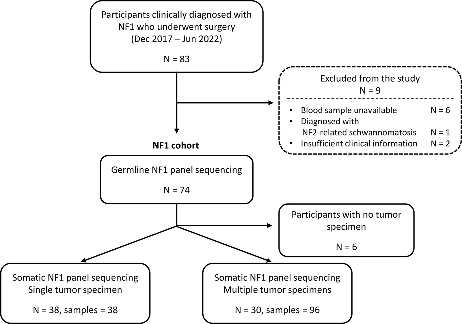
本研究最初纳入 83 例 2017 年 12 月至 2022 年 6 月间接受手术切除、临床诊断为 NF1 的个体(图 1)。8 例因不符合条件或无法获得血液样本被排除,1 例携带胚系 NF2 致病变异的患者被排除出 NF1 队列。最终,我们对 74 例患者的外周血样本进行了胚系 NF panel 测序(补充表 S2)。为识别体细胞突变,对 68 例患者的肿瘤标本进行了 panel 测序。其中 38 例患者有单份肿瘤标本,30 例患者共有 96 份肿瘤样本,系在单次手术中取自多个解剖部位,或来自不同时间的多次切除。6 例无可供分析肿瘤组织的患者仅接受胚系分析,未纳入体细胞谱分析(补充表 S3)。
各肿瘤样本的临床信息与组织病理学分类见补充表 S4。对 127 例 PNST 的组织病理学复核显示了显著的表型异质性,涵盖从良性神经纤维瘤到 MPNST 的完整组织学谱系(图 2)。肿瘤标本包括 61 例 CNF(其中 2 例为伴退变性非典型的神经纤维瘤)、26 例 PNF、15 例弥漫型神经纤维瘤、8 例混合型神经纤维瘤、9 例 ANNUBP、1 例低级别 MPNST 和 7 例高级别 MPNST。7 例因无法获得相应 H&E 切片而未能进行组织学复核。NF1 患者同时发生的其他肿瘤包括 3 例脑膜瘤、3 例 GIST、2 例神经内分泌肿瘤和 2 例乳腺癌。
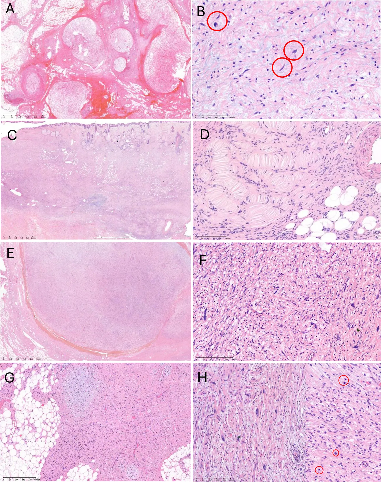
CNF 是最常见的亚型,特征为细胞密度低、梭形细胞温和、核呈波浪状。PNF 表现为疏松黏液样基质内扩张、迂曲的神经束,形成分叶状生长方式(图 2A)。若干病灶出现了不提示恶变前转化的少见组织学表现,包括神经纤维瘤中与退变性非典型相符的散在奇异核(图 2B),以及弥漫型神经纤维瘤中边界不清的真皮浸润(图 2C)。弥漫型神经纤维瘤可见类似 Meissner 小体的嗜酸性板层状结构(图 2D)24。
ANNUBP 表现为细胞密度增高(图 2E)、轻度核非典型(图 2F),非典型细胞中 p16 免疫组化表达灶性缺失(补充图 S1A、B),同时总体保留神经纤维瘤样结构。低级别 MPNST 部分保留原有结构,可见非典型梭形及上皮样细胞(图 2G),核分裂指数低,为 3−9 mf/10 HPF,无坏死(图 2H)。相比之下,高级别 MPNST 表现为致密的富细胞束状结构,伴显著细胞学非典型、坏死及活跃的核分裂(补充图 S1C、D)。这些发现展示了 NF1 相关 PNST 的组织病理学连续谱,并为下文所述的分子与组织病理学整合分析奠定了基础。
NF1 患者的胚系变异图谱
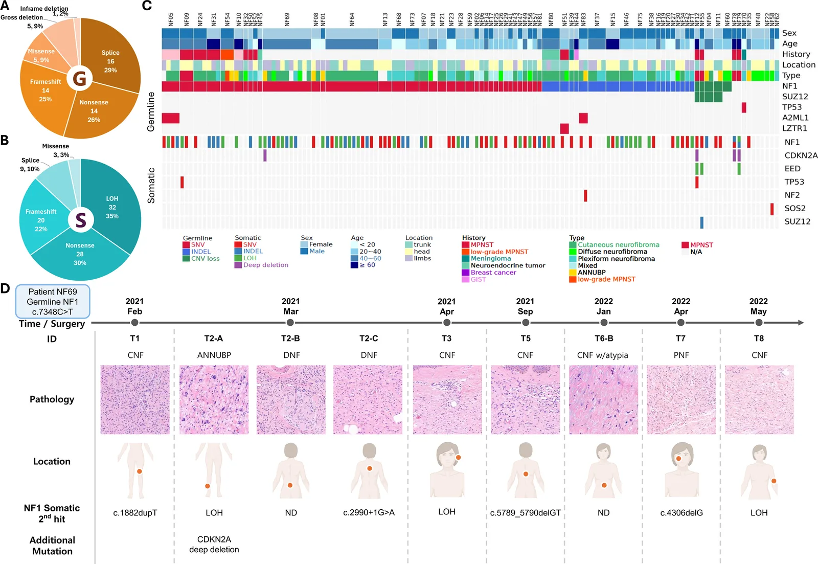
对 74 例临床诊断为 NF1 个体的血源 DNA 进行胚系变异分析,在 64 例患者(86.5%)中鉴定出 NF1 致病变异。值得注意的是,其中 17 例聚集为 8 个不同的家系,同一家系的受累成员携带相同的 NF1 变异,提示共同遗传。校正家系聚集后,我们共鉴定出 55 种不同的 NF1 变异(表 1)。在检出的 55 种非冗余变异中,21 种(47.7%)见于有阳性家族史的患者,23 种(52.3%)判定为新发(de novo);11 例因缺乏父母资料而无法核实。这与既往研究一致,即约半数受累个体的 NF1 源于新发的 NF1 致病变异7。值得注意的是,8 种变异(14.6%)为新变异,未见于 ClinVar 数据库(ClinVar 版本:2024-08-06)。在全部已鉴定的 NF1 致病变异中,最常见的分子亚型为剪接改变突变,占 55 种中的 16 种(29.1%)(图 3A)。除经典剪接位点破坏外,我们还在患者 NF26 中发现一个内含子深部变异,可引入隐蔽外显子(r.288_289ins288+1018_288+1135),导致移码,与既往报道的致病机制一致27。此外,两个外显子点突变 c.1885G>A(NF52)和 c.3304T>G(NF54)预测会在编码外显子内产生新的隐蔽剪接位点28,29,可能导致异常剪接。其余变异包括 14 个无义突变、14 个小 indel 所致移码突变、5 个错义突变、5 个大片段缺失和 1 个框内缺失。按 ACMG/AMP 指南,本队列全部变异均判定为致病或可能致病。另有一部分患者携带 NF1 微缺失,即累及 NF1 位点及其相邻基因组区域的大片段缺失。这类缺失通常由低拷贝重复序列之间的非等位同源重组介导。已描述两种复发性微缺失:1 型缺失(约 1.4 Mb),两侧为 NF1-REPa 和 NF1-REPc;2 型缺失(约 1.2 Mb),断点位于 SUZ12 及其假基因 SUZ12P 内30,31。在本队列中,拷贝数变异(CNV)分析鉴定出 5 个累及 NF1 基因的大片段缺失。患者 NF60 表现为外显子 8 内的局灶性缺失,导致外显子跳跃;其余 4 例则为累及整个 NF1 基因的更广泛 CNV 缺失。重要的是,这些患者的缺失边界各不相同,NF1 微缺失分别延伸至 SUZ12 外显子 3(NF04)、外显子 4(NF11)、外显子 8(NF12)以及整个 SUZ12 位点(NF55)(补充图 S2)。根据这些边界,4 个微缺失中有 3 个(75%)符合 2 型缺失,该比例高于既往报道的 11%32。在 4 例携带同时涵盖 NF1 与 SUZ12 的胚系大片段缺失的患者中,2 例(NF12 和 NF55)发生了高级别 MPNST,2 例发生了 ANNUBP,支持既往报道的 NF1 微缺失与恶性或恶变前病变终生风险升高相关31,33。
| NF1 (NM_001042492.2) | ||||||
|---|---|---|---|---|---|---|
| 病例 | 核苷酸改变 | 氨基酸改变 | 类型 | ClinVar 编号 | ClinVar 意义 | 遗传方式 |
| NF01 | c.8094C>A | p.Tyr2698* | 终止获得 | 963283 | P | 家族性 |
| NF02 | c.910C>T | p.Arg304* | 终止获得 | 187722 | P | 家族性 |
| NF03 | c.6642+1G>T | 外显子跳跃 | 剪接供体 | 404558 | P | NA |
| NF04 | WGD | - | - | 新发 | ||
| NF05 | c.5305C>T | p.Arg1769* | 终止获得 | 228381 | P | 新发 |
| NF06 | c.2332G>T | p.Glu778* | 终止获得 | 1434549 | P | 新发 |
| NF07 | c.1318C>T | p.Arg440* | 终止获得 | 230673 | P | 家族性 |
| NF08 | c.1527+1G>A | 外显子跳跃 | 剪接供体 | 503701 | P | 家族性 |
| NF10 | c.7348C>T | p.Arg2450* | 终止获得 | 185789 | P | 新发 |
| NF11 | WGD | - | - | 新发 | ||
| NF12 | WGD | - | - | NA | ||
| NF13 | c.217G>T | p.Glu73* | 终止获得 | 新变异 | - | 新发 |
| NF14 | c.7549C>T | p.Arg2517* | 终止获得 | 230467 | P | 家族性 |
| NF15 | c.5522_5523delAA | p.Gln1841fs | 移码 | 新变异 | - | 家族性 |
| NF16 | c.1756_1759delACTA | p.Thr586fs | 移码 | 2694619 | P | 家族性 |
| NF17 | c.968C>A | p.Ala323Asp | 错义 | 839743 | P | NA |
| NF18 | c.2288T>G | p.Leu763Arg | 错义 | 216396 | LP | 新发 |
| NF19 | c.591dupA | p.Ala198fs | 移码 | 新变异 | - | NA |
| NF20 | c.3525_3526delAA | p.Arg1176fs | 移码 | 428971 | P | 新发 |
| NF21 | c.479+1G>A | 外显子跳跃 | 剪接供体 | 457722 | P | 家族性 |
| NF23 | c.3198-2A>G | 外显子跳跃 | 剪接受体 | 428967 | P | NA |
| NF24 | c.4600C>T | p.Arg1534* | 终止获得 | 428967 | LP | 新发 |
| NF26# | c.288+1137C>T | p.Gln97Serfs | 诱导隐蔽外显子 | 2724584 | P | 家族性 |
| NF27 | c.499_502delTGTT | p.Cys167fs | 移码 | 185021 | P | 家族性 |
| NF28 | c.2851-2A>G | 外显子跳跃 | 剪接受体 | 2137973 | P | 新发 |
| NF29 | c.5768C>A | p.Thr1923Lys | 错义 | 945629 | P | 家族性 |
| NF31 | c.1721+3A>G | 外显子跳跃 | 剪接区域 | 374108 | P | 家族性 |
| NF32 | c.1260+1G>T | 外显子跳跃 | 剪接供体 | 570670 | LP | NA |
| NF34 | c.496_497delGT | p.Val166fs | 移码 | 431562 | P | 新发 |
| NF38 | c.2851-9_2851-5delTTTCT | 外显子跳跃 | 剪接区域 | 新变异 | - | 新发 |
| NF39 | c.4420dupG | p.Ala1474fs | 移码 | 1455367 | P | 新发 |
| NF41 | c.7909C>T | p.Arg2637* | 终止获得 | 184261 | LP | 新发 |
| NF42 | c.6852_6855delTTAC | p.Tyr2285fs | 移码 | 216866 | P | 家族性 |
| NF44 | c.4579delG | p.Asp1527fs | 移码 | 943771 | P | 新发 |
| NF45 | c.2339C>G | p.Thr780Arg | 错义 | 457581 | P | NA |
| NF46 | c.1872delT | p.Leu625fs | 移码 | 2137966 | P | 新发 |
| NF47 | c.2351G>A | p.Trp784* | 终止获得 | 2764008 | P | 新发 |
| NF49 | c.5503C>T | p.Gln1835* | 终止获得 | 852381 | P | 新发 |
| NF51 | c.5344_5345insG | p.Ile1782fs | 移码 | 新变异 | - | NA |
| NF52 | c.1885G>A | p.Gln616Glyfs | 新剪接位点 | 68308 | P | NA |
| NF54 | c.3304T>G | p.Leu1102Ilefs | 新剪接位点 | 1709572 | VUS | 新发 |
| NF55 | WGD | - | - | NA | ||
| NF56 | c.7189G>C | 外显子跳跃 | 错义及剪接区域 | 578365 | VUS | 家族性 |
| NF57 | c.6704+1G>C | 外显子跳跃 | 剪接供体 | 856149 | LP | 新发 |
| NF59 | c.7970+1G>C | 外显子跳跃 | 剪接供体 | 新变异 | - | 新发 |
| NF60 | 外显子 8 局灶性缺失 | - | - | 家族性 | ||
| NF64 | c.6819G>C | 外显子跳跃 | 错义及剪接区域 | 933505 | P | 家族性 |
| NF65 | c.288+3A>T | 外显子跳跃 | 剪接区域 | 942422 | VUS | 家族性 |
| NF71 | c.7184delT | p.Leu2395fs | 移码 | 1376674 | P | 家族性 |
| NF73 | c.3763C>T | p.Gln1255* | 终止获得 | 404504 | P | 家族性 |
| NF74 | c.5630T>C | p.Leu1877Pro | 错义 | 2113703 | P | 新发 |
| NF75 | c.1477_1481delCTCTT | p.Leu493fs | 移码 | 857230 | P | 新发 |
| NF80 | c.5857delC | p.Leu1953fs | 移码 | 新变异 | - | 家族性 |
| NF81 | c.2076C>G | p.Tyr692* | 终止获得 | 新变异 | - | 家族性 |
| NF83 | c.5036_5041delTCTATA | p.Ile1679_Tyr1680del | 框内缺失 | 431651 | LP | NA |
# 经 cDNA 测序检出,经病历复核发现;*,终止获得;fs,移码;NA,不详;WGD,全基因缺失;P,致病;LP,可能致病;VUS,意义未明变异。
NF1 患者肿瘤发生的遗传学特征
为探讨胚系遗传改变与 NF1 更广泛肿瘤谱之间的关系,我们将胚系测序结果与 PNST 及其他同时发生肿瘤的临床病理信息进行了整合(表 2)。共有 23 例患者被发现 NF1 相关的恶变前/恶性肿瘤或其他同时发生的肿瘤。这些肿瘤包括 15 例 MPNST(13 例发生于携带胚系 NF1 致病变异的患者,2 例发生于未检出胚系 NF1 变异的患者)、3 例 GIST、2 例乳腺癌、3 例脑膜瘤和 2 例神经内分泌肿瘤。这些发现凸显了 NF1 个体中所见肿瘤谱的多样性。GIST 和乳腺癌主要见于 ≥60 岁患者,而患者 NF05 在 60 岁前即发生 GIST,并同时携带 NF1 和 A2ML1 的致病变异;A2ML1 是已知与 RAS 病(RASopathies)相关的基因34。1 例儿童患者(NF70,<20 岁)表现为多发皮肤 CNF 和脑膜瘤。靶向 panel 检测未检出 NF1 致病变异,但测序发现一个胚系 TP53 无义突变(c.1175C>A;p.Ser392*)。Ser392 残基已被证实可通过磷酸化调节 TP53 的稳定性和转录活性35,36。尽管该变异此前未见与 NF1 表型相关的报道,但可能促成了本例多发肿瘤的发生。
| 病例 | 年龄 | NF 相关肿瘤* | 胚系致病变异 | NF1 突变类型 | 神经纤维瘤蛋白结构域# |
|---|---|---|---|---|---|
| NF03 | 40∼60 | MPNST | NF1 : c.6642+1G>T | 剪接 | 其他 |
| NF05 | 40∼60 | GIST | NF1 : p.Arg1769* ; A2ML1 : p.Gly637fs | 无义 | PH、LRD |
| NF09 | 40∼60 | MPNST | NF1 : p.Arg304* | 无义 | 其他 |
| NF10 | ≥ 60 | GIST、ANNUBP | NF1 : p.Arg2450* | 无义 | CTD |
| NF12 | ≥ 60 | MPNST、GIST | NF1 : CNV 缺失 ; SUZ12 : CNV 缺失 | 大片段缺失 | 全基因 |
| NF24 | 40∼60 | MPNST | NF1 : p.Arg1534* | 无义 | GRD |
| NF31 | ≥ 60 | CNS MPNST、乳腺癌、PNF | NF1 : c.1721+3A>G | 剪接 | CSRD |
| NF32 | 40∼60 | MPNST、DNF | NF1 : c.1260+1G>T | 剪接 | 其他 |
| NF39 | 40∼60 | CNS 脑膜瘤 | NF1 : p.Ala1474fs | 移码 | GRD |
| NF44 | ≥ 60 | 乳腺癌 | NF1 : p.Asp1527fs | 移码 | GRD |
| NF45 | ≥ 60 | 类癌、MNF | NF1 : p.Thr780Arg | 错义 | CSRD |
| NF50 | 20∼40 | MPNST、PNF | NF1 : c.1721+3A>G | 剪接 | CSRD |
| NF51 | 20∼40 | MPNST、DNF | NF1 : p.Ile1782fs ; LZTR1 : p.Arg412Cys | 移码 | PH、LRD |
| NF52 | < 20 | MPNST、PNF | NF1 : c.1885G>A | 剪接 | CSRD |
| NF54 | 40∼60 | 低级别 MPNST、ANNUBP | NF1 : c.3304T>G | 剪接 | TBD |
| NF55 | 20∼40 | MPNST | NF1 : CNV 缺失 ; SUZ12 : CNV 缺失 | 大片段缺失 | 全基因 |
| NF65 | 40∼60 | MPNST | NF1 : c.288+3A>T | 剪接 | 其他 |
| NF70 | < 20 | CNS 脑膜瘤、PNF | TP53 : p.Ser392* | ND | |
| NF76 | < 20 | MPNST | NF1 : p.Leu493fs | 移码 | 其他 |
| NF78 | ≥ 60 | MPNST | ND | ||
| NF79 | ≥ 60 | MPNST | ND | ||
| NF80 | 40∼60 | 嗜铬细胞瘤 | NF1 : p.Leu1953fs | 移码 | LRD |
* 本栏列出本研究所采集肿瘤的组织学类型,以及每例患者同时发生的 NF 相关肿瘤;皮肤神经纤维瘤未列出。PNF,丛状神经纤维瘤;DNF,弥漫型神经纤维瘤;MNF,混合型神经纤维瘤;CNS,中枢神经系统;ANNUBP,生物学潜能未定的非典型神经纤维瘤性肿瘤;MPNST,恶性周围神经鞘瘤。# 胚系 NF1 致病变异所累及的神经纤维瘤蛋白结构域。ND,未检出;CSRD,富半胱氨酸–丝氨酸结构域;TBD,微管蛋白结合结构域;GRD,GAP 相关结构域;PH,pleckstrin 同源结构域;LRD,富亮氨酸结构域;CTD,C 端结构域。
体细胞 NF1 突变在不同肿瘤部位与时间点间的克隆分化
我们分析了 68 例患者的 134 份肿瘤样本,以研究 NF1 相关肿瘤的体细胞突变图谱。配对血液样本中鉴定出的全部胚系 NF1 致病变异均在相应肿瘤组织中得到确认,表明胚系传递一致。此外,在携带配对胚系 NF1 变异的 124 个肿瘤中,有 90 个(72.6%)检出导致 NF1 双等位基因失活的体细胞突变。其中,LOH 是最常见的体细胞失活机制(图 3B)。值得注意的是,在 2 例无胚系 NF1 变异的 MPNST 中也观察到 NF1 双等位基因失活,进一步强调即便缺乏可检出的胚系易感性,NF1 缺失在 MPNST 发病中仍处于核心地位。
我们在恶变前及恶性病变中还发现了其他复发性体细胞改变。CDKN2A 纯合性拷贝数缺失见于 3 例 MPNST 和 1 例伴 p16 免疫组化表达缺失的 ANNUBP(补充图 S1A、B),与其在恶变前病变向恶性肿瘤进展中的作用相符37。此外,EED 的 LOH 以及 SUZ12 和 TP53 的体细胞突变仅见于 MPNST 和恶变前病变,而未见于良性神经纤维瘤(图 3C)。
为研究 NF1 患者肿瘤发生的肿瘤间异质性,我们分析了 30 例患者中多个解剖部位与时间上相互独立的肿瘤(补充表 S5)。图 3D 展示了一例代表性患者,其多份肿瘤样本取自不同解剖部位和时间点。患者 NF69 在共享胚系 NF1 致病变异(c.7348C>T)的背景下,突出显示了体细胞 NF1 改变的时空异质性。2021 年 2 月至 2022 年 5 月间共获得 11 个肿瘤,涵盖多种组织学亚型,包括 CNF、PNF、弥漫型神经纤维瘤、伴退变性非典型的皮肤神经纤维瘤以及 ANNUBP。各肿瘤样本的体细胞改变不同,几乎每个病灶都检出了不同的突变,包括 LOH、剪接位点变异(如 c.2990+1G>A)、小缺失(如 c.5789_5790delGT)和重复事件(如 c.1882dupT)。在级别较高的病灶(如 ANNUBP)中观察到共存改变,包括 NF1 的 LOH 和 CDKN2A 纯合缺失,提示其他抑癌基因的进一步破坏参与了疾病进展。
在大多数病例中,同一个体所发生的不同肿瘤之间体细胞 NF1 改变各不相同,提示其克隆起源在时间和空间上相互独立。尽管共享相同的胚系 NF1 变异,每个肿瘤都携带不同的体细胞变异,从而形成各自独立的双等位基因失活事件。仅 2 例患者(NF13 和 NF23)在全部取样肿瘤中携带相同的 NF1 变异;这 2 例均因解剖上连续的大病灶接受减瘤手术,提示所取组织很可能源自同一克隆。这些发现凸显了体细胞 NF1 失活作为 NF1 相关肿瘤发生中主要分子事件的关键作用,并进一步支持 NF1 中肿瘤以独立克隆方式发生——即在共同胚系易感背景下各自独立发生不同肿瘤。
基于显微切割的 WES 揭示恶性进展中的逐步分子演进
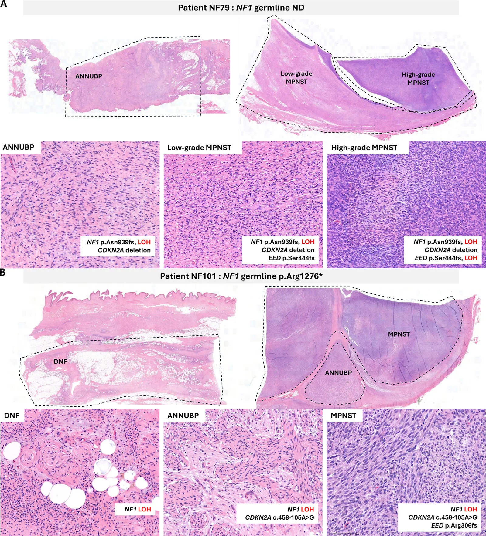
为进一步研究 NF1 相关 PNST 的瘤内分子演进,我们对 2 例代表性患者进行了基于显微切割的 WES(补充表 S6)。共对代表良性神经纤维瘤、ANNUBP、低级别 MPNST 和高级别 MPNST 成分的 6 个区域分别进行了分析(图 4)。患者 NF79 中,NF1 失活和 CDKN2A 缺失为各分析成分所共有,随后在低级别 MPNST 中获得 EED 突变,继而在高级别 MPNST 中出现 EED 杂合性缺失。患者 NF101 中,弥漫型神经纤维瘤、ANNUBP 和 MPNST 成分均存在 NF1 杂合性缺失,而 CDKN2A 改变出现于 ANNUBP 并在 MPNST 中保留,MPNST 中另检出 EED 突变。这些发现提示在恶性进展过程中 CDKN2A 及多梳抑制复合物 2(PRC2)通路被逐步破坏。
按组织学亚型的 NF1 变异结构域特异性分布
本队列所鉴定 NF1 变异的分布见图 5,并依据其在神经纤维瘤蛋白功能结构域中的定位,分别就胚系与体细胞突变进行了分析(补充图 S3)。两个热点 CSRD 和 CTD 呈胚系富集模式,但未达统计学意义(分别为 P = 0.08 和 P = 0.11),且胚系变异在各结构域间分布相对均匀。将 CSRD 与 CTD 合并后,相对于其他所有结构域呈显著的胚系富集(OR = 3.90,P = 0.006),并且在表现为多发 NF1 相关肿瘤或其他同时发生肿瘤的患者中,胚系 CSRD 与 CTD 变异频率略高(表 2)。相比之下,体细胞突变在另一热点 GRD 中最为富集(2.4 倍)(P = 0.008),而在 CTD 中代表性不足(0.25 倍)(补充图 S3C)。
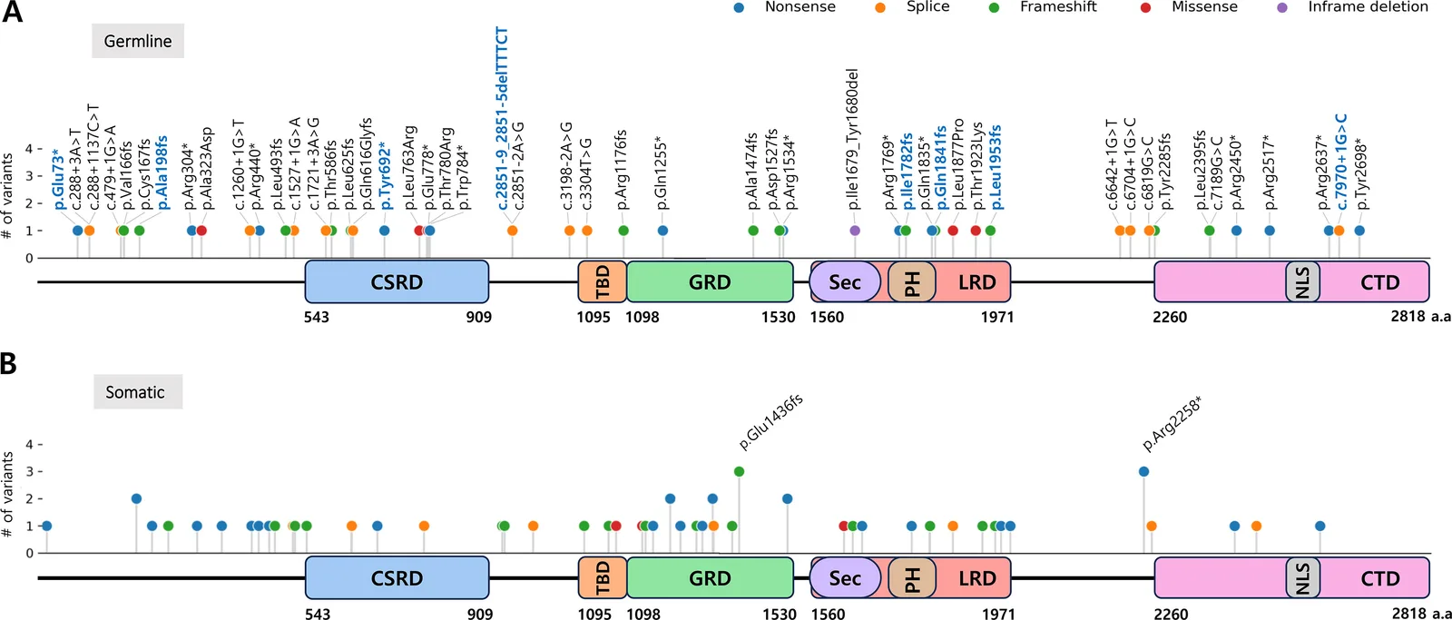
我们还将肿瘤组织学类型与突变累及的结构域位置相整合,汇总于表 3。重要的是,GRD 结构域在良性肿瘤中的突变频率最高(23.3% 对 0%,P = 0.035)(补充表 S7)。相比之下,含 Sec14-PH 亚结构域的 LRD 结构域变异在恶变前/恶性病变中较良性病变更常见(25.0% 对 8.2%),但差异未达统计学意义(P = 0.076)。此外,NF1 基因的 LOH 在所有组织学类型中均以相当比例出现。
| 良性 | 恶变前及恶性 | |||||||||
|---|---|---|---|---|---|---|---|---|---|---|
| 神经纤维瘤蛋白结构域* | 皮肤型 | 丛状 | 弥漫型 | 混合型 | MPNST | ANNUBP | NA | 合计 | 良性(%) | 其他(%) |
| CSRD | 3 | 0 | 0 | 1 | 0 | 0 | 0 | 4 | 4 (5.5) | 0 (0.0) |
| TBD | 1 | 0 | 0 | 1 | 0 | 1 | 0 | 3 | 2 (2.7) | 1 (6.3) |
| GRD | 8 | 8 | 1 | 0 | 0 | 0 | 0 | 17 | 17 (23.3) | 0 (0.0) |
| LRD (Sec-PH) | 5 | 0 | 0 | 1 | 1 | 3 | 1 | 11 | 6 (8.2) | 4 (25.0) |
| CTD | 2 | 0 | 0 | 0 | 1 | 0 | 0 | 3 | 2 (2.7) | 1 (6.3) |
| 其他区域 | 12 | 2 | 1 | 1 | 4 | 1 | 1 | 22 | 16 (21.9) | 5 (31.3) |
| LOH | 16 | 5 | 2 | 3 | 2 | 3 | 1 | 32 | 26 (35.6) | 5 (31.3) |
* 体细胞 NF1 致病变异所累及的神经纤维瘤蛋白结构域。CSRD,富半胱氨酸–丝氨酸结构域;TBD,微管蛋白结合结构域;GRD,GAP 相关结构域;Sec,Sec14 同源结构域;PH,pleckstrin 同源结构域;LRD,富亮氨酸结构域;CTD,C 端结构域;LOH,杂合性缺失;ANNUBP,生物学潜能未定的非典型神经纤维瘤性肿瘤;MPNST,恶性周围神经鞘瘤。
讨论
据我们所知,这是在空间与时间上相互独立的 NF1 相关 PNST 中整合胚系与体细胞改变、并对从良性 CNF 到高级别 MPNST 进行详细组织病理学对照的最大规模研究之一。
NF1 变异的结构域特异性定位显示,良性肿瘤的体细胞突变富集于 GRD,提示其可能在早期肿瘤发生中发挥作用;而 LRD(Sec14-PH)结构域变异在恶变前及恶性病变中呈富集趋势,但未达统计学意义。鉴于本队列中恶变前与恶性肿瘤数量有限,需更大规模研究来确定结构域特异性 NF1 改变是否促成恶性进展。从治疗角度看,这些发现提示 RAS/MAPK 通路抑制可能对 PNF 等良性病变尤为相关;相比之下,恶性进展可能需要 MEK 抑制之外的其他治疗策略。多发或同时发生肿瘤的患者中胚系 CSRD 与 CTD 变异频率略高,提示其可能与更广泛的肿瘤易感性相关,但这一观察有待更大队列验证。
对同一个体多个肿瘤的分析显示出显著的体细胞异质性:不同肿瘤在共同胚系背景下独立发生。这一发现支持个体内各肿瘤之间存在病灶特异性的分子分化,而非共同的体细胞驱动事件。多种恶性及良性肿瘤——包括 MPNST、GIST、乳腺癌、脑膜瘤和神经内分泌肿瘤——的发生,凸显了 NF1 相关肿瘤易感性的多系统特性。
我们的结果强调了 NF1 失活在 PNST 发病中的关键作用20,38。与既往发现一致,涵盖 SUZ12 的 NF1 微缺失与恶性潜能升高相关33。在携带同时涵盖 NF1 与 SUZ12 的胚系缺失的患者中观察到 MPNST 和 ANNUBP,与既往关于大片段缺失使恶性或恶变前转化风险升高的证据一致。在 1 例 NF1 panel 阴性的儿童病例中鉴定出胚系 TP53 无义变异,提示在部分患者中继发性遗传事件可能参与肿瘤发生。此外,高级别病变中调控染色质重塑(SUZ12、EED)和细胞周期(CDKN2A、TP53)的基因同时发生体细胞改变,凸显了 NF1 相关肿瘤发生的多步骤性质。本研究中基于显微切割的 WES 进一步展示了从 NF1 相关神经纤维瘤到 MPNST 的组织学不同成分间的分子演进。值得注意的是,在较晚期的肿瘤成分中鉴定出累及 PRC2 核心组分 EED 的额外改变,支持其在进展过程中获得。总体而言,这些数据支持以下观点:NF1 失活与其他体细胞或胚系改变协同作用,共同塑造 NF1 个体的肿瘤谱和临床结局。
与既往主要关注胚系变异或单一肿瘤类型的研究不同,本研究整合了时空上相互独立的肿瘤在多种组织学亚型中的胚系与体细胞数据,揭示了显著的患者内与患者间异质性。根据近期一项基因型优先的研究39,NF1 致病变异的患病率可能高于既往估计。它们可见于缺乏经典临床特征的个体,常表现为体细胞嵌合或外显率降低。与这些发现一致,更新的遗传学诊断标准已被提出,并强烈共识推荐对有节段性表现的患者以及有多发双侧脊神经神经纤维瘤但无其他 NF1 特征的个体进行检测18。我们鉴定出 8 种既往未报道的胚系变异,扩展了已知的 NF1 突变谱,可能有助于改进分子诊断。
本研究存在一些局限性。第一,单中心设计可能限制结果的普适性。虽然就 NF1 相关 PNST 研究而言本队列规模相对较大,但恶变前病变、MPNST 及多灶样本的数量有限。第二,我们认识到,近期更新的 NF1 相关肿瘤国际共识推荐40已不再支持使用“低级别 MPNST”这一术语。本队列中 1 例最初依据 2020 年 WHO 分类及当时可用的病理标准诊断为低级别 MPNST。为与历史分类保持一致,我们保留了原诊断,同时认识到此类病变按现行共识推荐可能会被归入不同类别。第三,尽管我们的 62 基因靶向 panel 对 NF1 相关肿瘤发生通路覆盖全面,但仍可能遗漏 panel 之外的其他基因组改变。我们未对所鉴定的变异进行功能验证,panel 之外的非编码或结构变异可能在 panel 阴性病例中影响肿瘤表型。最后,本队列中无胚系 NF1 致病变异的 MPNST 过少,无法与 NF1 相关 MPNST 进行有意义的比较;因此,我们未能直接比较 NF1 相关与散发性 MPNST 的分子谱。今后需纳入两组更多病例的研究,以界定其共有及不同的基因组特征。
总之,我们的发现具有若干重要的临床意义。第一,体细胞 NF1 变异的结构域特异性富集若在更大队列中得到验证,可为基因型–表型相关提供依据并有助于风险分层。第二,病灶特异性的分子分化凸显了在制定个体化治疗策略时进行病灶特异性基因组谱分析的必要性,尤其是对多发或高级别肿瘤患者。第三,恶性进展过程中染色质重塑与细胞周期调控基因改变的逐步获得,提示在高危 NF1 相关肿瘤中存在 MEK 抑制之外的潜在治疗靶点。今后需开展纳入空间分辨基因组学方法及更广泛测序策略(包括全基因组测序)的多中心研究,以验证基因型–表型相关并识别其他肿瘤发生驱动因素,包括非编码和结构变异。这些认识可为开发将 RAS/MAPK 通路抑制剂与其他药物联合、针对高危 NF1 相关肿瘤的新型治疗方法提供指导。
- 74 NF1 patients, 134 tumours: germline + somatic 62-gene panel sequencing with histological review per the 2016 NCI consensus and 2020 WHO (CNF, PNF, diffuse-type, ANNUBP, low/high-grade MPNST).
- Biallelic NF1 inactivation in 72.6% of tumours, most often by LOH; CDKN2A homozygous loss, EED LOH and SUZ12/TP53 mutations only in ANNUBP/MPNST, not benign neurofibromas.
- Separate tumours in one patient usually carried distinct NF1 second hits; components within one tumour (ANNUBP→MPNST) shared NF1 alterations and sequentially acquired CDKN2A and EED changes.
- Of 4 patients with germline NF1 microdeletions involving SUZ12, 2 developed high-grade MPNST and 2 ANNUBP.
Scope: open-access full text (CC BY 4.0) from Modern Pathology, including 5 figures and 3 tables; supplementary material not included. Superscript numbers are the original reference numbers; see the original article for the reference list.
Abstract
Peripheral nerve sheath tumors (PNSTs) arising in neurofibromatosis type 1 (NF1) show marked histological and clinical heterogeneity, ranging from benign to malignant PNSTs. Although germline NF1 variants underlie tumor predisposition, the mechanisms driving phenotypic and clinical heterogeneity remain incompletely defined. We performed integrated germline and somatic analyses of 208 samples (74 blood samples and 134 tumors) from a primary cohort of 74 patients with NF1. Using a custom NF-focused sequencing panel, we investigated NF1 variant types, co-occurring oncogenic alterations, and affected domains to assess genotype–phenotype associations. Germline analysis identified 55 pathogenic NF1 variants, including eight unreported variants, with recurrent NF1 microdeletions involving SUZ12 in high-grade and premalignant tumors. Among tumors from individuals with germline NF1 variants, biallelic NF1 inactivation occurred in 90/124 tumors (72.6%), most frequently through loss of heterozygosity. In addition, high-grade PNSTs frequently harbored co-alterations in CDKN2A, SUZ12, EED, or TP53. Analysis of the distribution of somatic NF1 variants across functional domains further revealed significant enrichment in the GRD in benign tumors, whereas variants involving the LRD, including the Sec14-PH subdomain, showed a trend toward enrichment in premalignant and malignant tumors. Notably, multi-lesion analysis of spatially and temporally distinct tumors from 30 individuals revealed pronounced intrapatient somatic heterogeneity: despite a shared germline NF1 background, individual tumors typically carried distinct NF1 second-hit events, consistent with lesion-specific molecular divergence rather than a single shared somatic driver across lesions. In contrast, microdissected histologically distinct components within individual tumors shared underlying NF1 alterations and showed sequential acquisition of additional alterations involving CDKN2A and EED, supporting stepwise molecular evolution during malignant progression. Together, these data reveal lesion-specific molecular divergence across distinct tumors and stepwise molecular evolution within individual tumors, underscoring the value of lesion-specific genomic evaluation in NF1-associated PNSTs.
Introduction
Neurofibromatosis (NF) syndromes are characterized by multiple neural tumors, particularly Schwann cell neoplasms, which manifest in different locations 1. These syndromes include neurofibromatosis type 1 (NF1) and schwannomatosis. Among them, NF1 is an autosomal dominant genetic disorder with an estimated incidence of approximately 1 in 3,000 individuals worldwide 2-4. It is caused by pathogenic variants in the NF1 gene, a tumor suppressor gene located on chromosome 17q11.2. 5,6.
NF1 is a multisystem disorder characterized by significant phenotypic variability, ranging from benign peripheral nerve sheath tumors (PNSTs) to more severe malignancies, including malignant peripheral nerve sheath tumors (MPNSTs) 7. Due to the systemic involvement and tumor-suppressive function of the NF1 gene, individuals with NF1 are prone to developing diverse tumor types, such as breast cancer, gastrointestinal stromal tumors (GISTs), and neuroendocrine neoplasms 8.
NF1-associated PNSTs encompass a wide range of cutaneous neurofibromas (CNFs), plexiform neurofibromas (PNFs), atypical neurofibromatous neoplasms of uncertain biologic potential (ANNUBP), and MPNSTs 9. Nearly all NF1 patients develop CNFs, which are confined to cutaneous tissue and may number in the hundreds or thousands 10. Unlike CNFs, the surgical removal of PNFs is challenging because PNFs occur deeper and involve multiple nerve bundles. The FDA approved the MAPK/ERK pathway (MEK) inhibitor, selumetinib, for the treatment of children with symptomatic inoperable PNF, based on a clinical trial demonstrating significant tumor volume reduction in children with NF1 1,11. However, MEK inhibitors are not curative, and PNFs carry a risk of malignant transformation to MPNSTs 7. Approximately 8–13% of individuals with NF1 develop MPNSTs during their lifetime 12. Because of their highly aggressive behavior, MPNSTs are treated by surgical resection with wide margins 13, radiation, and chemotherapy in a primary or adjuvant setting. Despite these approaches, the local recurrence rates remain high (∼32–65%), highlighting an urgent need for more effective treatments 14.
NF1 is caused by pathogenic variants in the NF1 gene, which encodes neurofibromin, a tumor suppressor protein that negatively regulates the RAS/MAPK signaling pathway. More than 3,000 disease-causing NF1 variants have been reported 15, contributing to the marked genetic and clinical heterogeneity of the disease. These variants include nonsense, frameshift, splice-site, and missense variants, as well as multi-exonic deletions, which can reduce neurofibromin function. Neurofibromin harbors multiple functional domains that mediate interactions with various cellular components, potentially contributing to the diverse clinical manifestations of NF1 16,17. Recently, clinically relevant genotype–phenotype correlations have been identified 18,19, such as an association between NF1 microdeletions and MPNST development and three hotspot regions within neurofibromin, including the GAP-related domain (GRD, residues 1198–1530), cysteine/serine-rich domain (CSRD, residues 543–909), and Armadillo1 domain within the C-terminal domain (CTD, residues 2260–2818) 20. These findings suggest that the type and location of NF1 variants may contribute to the phenotypic heterogeneity of NF1-associated tumors.
Despite substantial progress in germline genetic testing for NF1 diagnosis, the molecular mechanisms underlying the remarkable histological and clinical heterogeneity of PNSTs in NF1 remain incompletely understood. Although a recent study suggested that somatic second-hit events may occur independently in spatially distinct lesions in NF1 21, whether multiple tumors arising within an individual or among members of the same family share common somatic drivers or develop independently has not been systematically investigated. Furthermore, correlations between the histopathology of PNSTs and genetic alterations remain underexplored, aside from a limited number of established genotype–phenotype associations. Thus, beyond advances in diagnostic genetics, comprehensive, systematic acquisition of human tumor specimens, coupled with integrated molecular analyses, is essential to delineate disease mechanisms and facilitate the development of effective therapies for NF1-associated PNSTs 9. In this study, we performed integrated germline and somatic analyses in patients with clinically diagnosed NF1, combining genomic profiling with detailed histopathological classification. This approach allowed us to characterize domain-specific patterns of NF1 alterations and to demonstrate that NF1-associated PNSTs develop through divergent molecular pathways, despite a shared germline predisposition. Our findings provide further insight into NF1-associated tumorigenesis and highlight the therapeutic implications of lesion-specific genomic heterogeneity.
Materials & Methods
Patient Sample Collection
Participants clinically diagnosed with NF1 who underwent surgery at the Samsung Medical Center (SMC) in Seoul, Korea, between December 2017 and June 2022 were enrolled. A total of 74 blood samples and 134 tissue samples were subjected to targeted sequencing. Immediately after surgery, a portion of each resected specimen was separated by the surgeon in the operating room and stored at –80 °C as fresh-frozen tissue for NGS analysis. The remaining tissue was submitted to the Department of Pathology and processed into formalin-fixed paraffin-embedded (FFPE) blocks for histopathological evaluation. The histopathological diagnoses were confirmed using H&E-stained sections prepared from the corresponding FFPE tissue obtained from the same surgical specimen. Additional microdissection-based whole-exome sequencing (WES) was performed on six histologically distinct regions from two patients: one patient from the primary cohort and one additional patient who was included exclusively in the exploratory microdissection-based WES analysis. All studies were approved by the SMC Institutional Review Board (IRB Nos. 2013-09-025 and 2026-08-095), and written informed consent was obtained from all patients in the primary cohort. The requirement for informed consent was waived by the IRB for one additional patient who was retrospectively included in the study.
Pathology Review
Clinical and radiological data were extracted from patients' medical records. The collected clinical information included age, sex, tumor location, date of excision, family history, associated symptoms, medical history, computed tomography and magnetic resonance imaging findings, follow-up duration, treatment history, and pathological diagnosis. Two board-certified pathologists reviewed all available slides and classified the tumors according to the proposed nomenclature from the consensus meeting on the pathology of NF1-associated atypical nerve sheath tumors held in October 2016 at the National Cancer Institute/National Institutes of Health 22 and the 2020 World Health Organization (WHO) classification of bone and soft tissue tumors 23. We classified NF1-associated nerve sheath tumors into the following categories: CNF, PNF, diffuse-type neurofibroma, neurofibroma with degenerative atypia, ANNUBP, low- and high-grade MPNST. CNFs were defined as benign, localized dermal neurofibromas. PNFs involved multiple nerve fascicles and exhibited a tortuous, multinodular growth pattern. Neurofibromas showing nuclear atypia, without hypercellularity or increased mitosis, were termed neurofibroma with degenerative atypia. Tumors exhibiting at least two of the four features—such as cytologic atypia, loss of neurofibroma architecture, hypercellularity, and a mitotic index >1/10 and < 3/10 high-power fields (HPFs)—were classified as ANNUBP. Low-grade MPNST (or ANNUBP with increased proliferation) showed ANNUBP features, but with mitotic figures (mf) of 3–9/10 HPFs and no necrosis. If an MPNST had 3–9 mf/10 HPFs with necrosis or at least 10 mf/10 HPFs, it was classified as a high-grade MPNST. Diffuse-type neurofibromas were retained as a separate subtype because they can clinically mimic large malignant tumors but exhibit histological features distinct from classical neurofibromas 24,25. Tumors exhibiting both the plexiform and diffuse growth patterns were classified as mixed-type neurofibromas. In addition, slides from other concurrent tumors occurring in patients with NF1 were reviewed to confirm pathological diagnoses.
NF-panel Design and NGS Sequencing
To investigate genetic alterations in NF1-associated tumors, we designed a custom-targeted sequencing panel comprising 62 genes (Supplementary Table S1). Genes were selected based on their known involvement in the tumorigenesis of NF-associated tumors, including core components of the RAS/MAPK pathway7,26 (e.g., NF1, NRAS, KRAS, HRAS, MAP2K1/2), cell cycle regulation (CDKN2A, TP53, CCND2), chromatin remodeling (SUZ12, EED, CHD4, SMARCB1), and other tumor suppressors or oncogenes that are frequently altered in peripheral nerve sheath tumors. Celemics Inc. (Seoul, Korea) manufactured the panel as a custom target-capture panel. Capture probes were designed to cover all coding exons of selected genes and the 50-bp intronic regions adjacent to exon boundaries. DNA was extracted from fresh-frozen tissue samples using the QIAamp DNA Mini Kit (Qiagen, Hilden, Germany) and from blood samples using the QIAamp DNA Blood Maxi Kit (Qiagen). Library preparation followed standard enzymatic steps (end repair, A-tailing, adapter ligation, and PCR amplification) using a Celemics Library Preparation Kit (Celemics, Seoul, Korea). DNA libraries were hybridized with capture probes in buffer using a Celemics Target Enrichment Kit (Celemics, Korea). After hybridization and washing, the captured libraries underwent post-capture PCR amplification. Pooled libraries were sequenced on an Illumina NextSeq 500 Sequencing System (Illumina, San Diego, CA, USA) using 2 × 151-bp paired-end reads.
Bioinformatics Analysis of Targeted Sequencing Data
Raw FASTQ files were processed using AdapterRemoval and aligned to the human reference genome (GRCh37) using BWA-MEM. Duplicate reads were removed using Picard, and subsequent sequence processing was performed using GATK. Copy number variation (CNV) values were calculated by dividing the normalized regional depth of the test sample by the average normalized regional depth of the control samples. Short-base variants were identified using VarDict and annotated using SnpEff based on their genomic locations and predicted coding effects. We analyzed genomic variants, including SNPs, INDELs, MNPs, CNVs, and translocations, for clinical significance using the ClinVar database. Germline variants with a population frequency of <0.1% were reviewed, and somatic variants with variant allele frequency (VAF) >5% were selected. Loss of heterozygosity (LOH) was determined by an increase in the VAF of the germline pathogenic variant in tumor samples compared to that in matched blood samples.
Whole-exome Sequencing and Bioinformatics Analysis of Microdissected FFPE Samples
Additional WES was performed on six histologically distinct regions from two patients, including diffuse-type neurofibroma, ANNUBP, low-grade MPNST, and high-grade MPNST. The corresponding regions were manually microdissected from unstained FFPE tissue sections. Exome libraries were prepared from 100 ng of genomic DNA using the Agilent SureSelect XT HS/Low Input protocol and SureSelect Human All Exon V8 kit and sequenced with 151-bp paired-end reads by Macrogen (Seoul, Korea). After adapter trimming with AGeNT Trimmer (version 3.0.6), reads were aligned to the GRCh38 human reference genome using BWA-MEM (version 0.7.17). Molecular barcode–based duplicate processing and base quality score recalibration were subsequently performed. Somatic SNVs and small insertions/deletions were called using GATK Mutect2 in tumor-normal paired mode and annotated using SnpEff (version 5.2).
Statistical Analysis
Oncoprint visualization was generated using the PyComplexHeatmap (version 1.7.4) in Python. Graphical analyses were conducted using Seaborn (version 0.11.0), and schematic figures were created using BioRender.com. The frequency of domain distribution was evaluated using Fisher's exact test. A two-sided P value less than 0.05 was considered statistically significant. Statistical analyses were performed using Rex 3.6.0, an Excel-based statistical analysis software package (RexSoft, Korea; http://rexsoft.org/) and R ver. 4.0.0 (R Foundation for Statistical Computing, Austria).
Results
Sample Collection and Histological Classification for Targeted Sequencing
A total of 83 individuals with a clinical diagnosis of NF1 who underwent surgical resection between December 2017 and June 2022 were initially enrolled for this study (Fig. 1). Eight patients were excluded because of ineligibility or the unavailability of blood samples, and one patient harboring a germline NF2 pathogenic variant was excluded from the NF1 cohort. Finally, we performed germline NF panel sequencing on peripheral blood samples from 74 patients. (Supplementary Table S2). To identify somatic mutations, tumor specimens from 68 patients were subjected to panel sequencing. Among them, 38 patients had a single tumor specimen, and 30 patients had a total of 96 tumor samples obtained either from multiple anatomical sites during a single surgical procedure or from serial resections over time. Six patients without tumor tissue available for analysis underwent germline analysis alone and were excluded from somatic profiling (Supplementary Table S3).
The clinical information and histopathological classification of each tumor sample are provided in Supplementary Table S4. A histopathological review of 127 PNST cases demonstrated marked phenotypic heterogeneity, encompassing the full histological spectrum from benign neurofibromas to MPNSTs (Fig. 2). The tumor specimens included 61 CNFs, including two neurofibromas with degenerative atypia, 26 PNFs, 15 diffuse-type neurofibromas, eight mixed-type neurofibromas, nine ANNUBP, one low-grade MPNST, and seven high-grade MPNSTs. Seven cases could not be histologically reviewed because the corresponding H&E slides were unavailable. Other concurrent tumors in patients with NF1 included three meningiomas, three GISTs, two neuroendocrine tumors, and two breast carcinomas.
CNFs were the most prevalent subtype, characterized by low cellularity and bland spindle cells with wavy nuclei. PNFs exhibited expanded tortuous nerve fascicles within a loose myxoid matrix, resulting in a lobulated growth pattern (Fig. 2A). Several lesions showed unusual histological findings not indicative of premalignant transformation, including scattered bizarre nuclei consistent with degenerative atypia in neurofibromas (Fig. 2B) and poorly circumscribed dermal infiltration in diffuse-type neurofibromas (Fig. 2C). Diffuse-type neurofibromas exhibited eosinophilic lamellar structures resembling Meissner corpuscles (Fig.2D) 24.
ANNUBPs exhibited increased cellularity (Fig. 2E), mild nuclear atypia (Fig. 2F), and focal loss of p16 immunohistochemistry expression in atypical cells (Supplementary Fig. S1A, B), while retaining an overall neurofibromatous architecture. Low-grade MPNSTs showed partial preservation of the underlying architecture with atypical spindle and epithelioid cells (Fig. 2G) and a low mitotic index of 3−9 mf/10 HPFs without necrosis (Fig. 2H). In contrast, high-grade MPNSTs demonstrated dense cellular fascicles with marked cytological atypia, necrosis, and brisk mitotic activity (Supplementary Fig. S1C, D). These findings illustrate the histopathological continuum of NF1-associated PNSTs and provide a basis for integrating molecular and histopathological analyses described below.
Germline Variant Landscape of NF1 Patients
Germline variant analysis was performed on blood-derived DNA from 74 individuals clinically diagnosed with NF1, identifying pathogenic variants in NF1 in 64 patients (86.5%). Notably, 17 cases were clustered into eight distinct familial groups, with affected members of each family harboring the same NF1 variant, indicating shared inheritance. After accounting for familial clustering, we identified 55 distinct NF1 variants (Table 1). Among the 55 non-redundant variants detected, 21 (47.7%) were identified in patients with a positive family history, whereas 23 (52.3%) were classified as de novo; 11 cases remained unverifiable owing to a lack of parental data. This is consistent with a previous study, which found that approximately half of affected individuals had NF1 due to a de novo NF1 disease-causing variant 7. Of note, eight variants (14.6%) were novel and had not been reported in the ClinVar database (ClinVar release: 2024-08-06). Among all identified pathogenic NF1 variants, the most frequent molecular subtype was splice-altering mutations, which accounted for 16 of 55 variants (29.1%) (Fig. 3A). In addition to canonical splice site disruptions, we identified a deep intronic variant in patient NF26, which introduced a cryptic exon (r.288_289ins288+1018_288+1135), resulting in a frameshift, consistent with a previously reported pathogenic mechanism 27. Moreover, two exonic point mutations, c.1885G>A (NF52) and c.3304T>G (NF54), were predicted to generate novel cryptic splice sites within coding exons28,29, likely leading to aberrant splicing. The remaining variants comprised 14 nonsense mutations, 14 small indel-induced frameshift mutations, five missense mutations, five large deletions, and one in-frame deletion. All variants in this cohort were classified as pathogenic or likely pathogenic according to the ACMG/AMP guidelines. A distinct subset of patients harbored NF1 microdeletions, defined as large deletions involving the NF1 locus and its adjacent genomic regions. These deletions are typically mediated by nonallelic homologous recombination between low-copy repeats. Two recurrent types of microdeletions have been described: type-1 deletion (∼1.4 Mb), flanked by NF1-REPa and NF1-REPc, and type-2 deletion (∼1.2 Mb), with breakpoints located within SUZ12 and its pseudogene SUZ12P 30,31. In our cohort, copy number variation (CNV) analysis identified five large deletions involving the NF1 gene. Patient NF60 exhibited a focal deletion within exon 8, leading to exon skipping, whereas the remaining four patients showed broader CNV losses affecting the entire NF1 gene. Importantly, deletion boundaries varied among these patients, with NF1 microdeletions extending to SUZ12 exons 3 (NF04), 4 (NF11), 8 (NF12), and the entire SUZ12 locus (NF55) (Supplementary Fig. S2). Based on these boundaries, three of the four microdeletions (75%) were consistent with type-2 deletions, a proportion higher than the previously reported frequency of 11% 32. Among the four patients carrying large germline deletions encompassing both NF1 and SUZ12, two (NF12 and NF55) developed high-grade MPNSTs, and two developed ANNUBPs, supporting the elevated lifetime risk of malignant or premalignant lesions associated with NF1 microdeletions, as previously reported 31,33.
| NF1 (NM_001042492.2) | ||||||
|---|---|---|---|---|---|---|
| Case | Nucleotide change | Amino acid change | Type | ClinVar ID | ClinVar significance | Inherit. |
| NF01 | c.8094C>A | p.Tyr2698* | stop-gain | 963283 | P | familial |
| NF02 | c.910C>T | p.Arg304* | stop-gain | 187722 | P | familial |
| NF03 | c.6642+1G>T | exon skip | splice donor | 404558 | P | NA |
| NF04 | WGD | - | - | de novo | ||
| NF05 | c.5305C>T | p.Arg1769* | stop-gain | 228381 | P | de novo |
| NF06 | c.2332G>T | p.Glu778* | stop-gain | 1434549 | P | de novo |
| NF07 | c.1318C>T | p.Arg440* | stop-gain | 230673 | P | familial |
| NF08 | c.1527+1G>A | exon skip | splice donor | 503701 | P | familial |
| NF10 | c.7348C>T | p.Arg2450* | stop-gain | 185789 | P | de novo |
| NF11 | WGD | - | - | de novo | ||
| NF12 | WGD | - | - | NA | ||
| NF13 | c.217G>T | p.Glu73* | stop-gain | novel | - | de novo |
| NF14 | c.7549C>T | p.Arg2517* | stop-gain | 230467 | P | familial |
| NF15 | c.5522_5523delAA | p.Gln1841fs | frameshift | novel | - | familial |
| NF16 | c.1756_1759delACTA | p.Thr586fs | frameshift | 2694619 | P | familial |
| NF17 | c.968C>A | p.Ala323Asp | missense | 839743 | P | NA |
| NF18 | c.2288T>G | p.Leu763Arg | missense | 216396 | LP | de novo |
| NF19 | c.591dupA | p.Ala198fs | frameshift | novel | - | NA |
| NF20 | c.3525_3526delAA | p.Arg1176fs | frameshift | 428971 | P | de novo |
| NF21 | c.479+1G>A | exon skip | splice donor | 457722 | P | familial |
| NF23 | c.3198-2A>G | exon skip | splice acceptor | 428967 | P | NA |
| NF24 | c.4600C>T | p.Arg1534* | stop-gain | 428967 | LP | de novo |
| NF26# | c.288+1137C>T | p.Gln97Serfs | cryptic exon induction | 2724584 | P | familial |
| NF27 | c.499_502delTGTT | p.Cys167fs | frameshift | 185021 | P | familial |
| NF28 | c.2851-2A>G | exon skip | splice acceptor | 2137973 | P | de novo |
| NF29 | c.5768C>A | p.Thr1923Lys | missense | 945629 | P | familial |
| NF31 | c.1721+3A>G | exon skip | splice region | 374108 | P | familial |
| NF32 | c.1260+1G>T | exon skip | splice donor | 570670 | LP | NA |
| NF34 | c.496_497delGT | p.Val166fs | frameshift | 431562 | P | de novo |
| NF38 | c.2851-9_2851-5delTTTCT | exon skip | splice region | novel | - | de novo |
| NF39 | c.4420dupG | p.Ala1474fs | frameshift | 1455367 | P | de novo |
| NF41 | c.7909C>T | p.Arg2637* | stop-gain | 184261 | LP | de novo |
| NF42 | c.6852_6855delTTAC | p.Tyr2285fs | frameshift | 216866 | P | familial |
| NF44 | c.4579delG | p.Asp1527fs | frameshift | 943771 | P | de novo |
| NF45 | c.2339C>G | p.Thr780Arg | missense | 457581 | P | NA |
| NF46 | c.1872delT | p.Leu625fs | frameshift | 2137966 | P | de novo |
| NF47 | c.2351G>A | p.Trp784* | stop-gain | 2764008 | P | de novo |
| NF49 | c.5503C>T | p.Gln1835* | stop-gain | 852381 | P | de novo |
| NF51 | c.5344_5345insG | p.Ile1782fs | frameshift | novel | - | NA |
| NF52 | c.1885G>A | p.Gln616Glyfs | new splice site | 68308 | P | NA |
| NF54 | c.3304T>G | p.Leu1102Ilefs | new splice site | 1709572 | VUS | de novo |
| NF55 | WGD | - | - | NA | ||
| NF56 | c.7189G>C | exon skip | missense&splice region | 578365 | VUS | familial |
| NF57 | c.6704+1G>C | exon skip | splice donor | 856149 | LP | de novo |
| NF59 | c.7970+1G>C | exon skip | splice donor | novel | - | de novo |
| NF60 | exon 8 focal deletion | - | - | familial | ||
| NF64 | c.6819G>C | exon skip | missense&splice region | 933505 | P | familial |
| NF65 | c.288+3A>T | exon skip | splice region | 942422 | VUS | familial |
| NF71 | c.7184delT | p.Leu2395fs | frameshift | 1376674 | P | familial |
| NF73 | c.3763C>T | p.Gln1255* | stop-gain | 404504 | P | familial |
| NF74 | c.5630T>C | p.Leu1877Pro | missense | 2113703 | P | de novo |
| NF75 | c.1477_1481delCTCTT | p.Leu493fs | frameshift | 857230 | P | de novo |
| NF80 | c.5857delC | p.Leu1953fs | frameshift | novel | - | familial |
| NF81 | c.2076C>G | p.Tyr692* | stop-gain | novel | - | familial |
| NF83 | c.5036_5041delTCTATA | p.Ile1679_Tyr1680del | inframe deletion | 431651 | LP | NA |
# Detected by cDNA sequencing, found through medical chart review; *, stop gain; fs, frameshift, NA, not available; WGD, whole gene deletion; P, pathogenic; LP, likely pathogenic; VUS, variant of uncertain significance
Genetic Features Underpinning Tumor Development in NF1 Patients
To explore the relationship between germline genetic alterations and the broader tumor spectrum in NF1, we integrated germline sequencing results with the clinicopathologic information of PNSTs and other concurrent tumors (Table 2). In total, NF1-associated premalignant/malignant tumors or other concurrent tumors were identified in 23 patients. These tumors included 15 MPNSTs (13 MPNSTs arising in patients with germline NF1 pathogenic variants and two MPNSTs arising in patients without detectable germline NF1 variants), three GISTs, two breast cancers, three meningiomas, and two neuroendocrine tumors. These findings underscore the diverse tumor spectrum observed in individuals with NF1. Whereas GIST and breast cancer mainly occurred in patients aged ≥60 years, patient NF05 developed a GIST before 60 years of age and carried pathogenic variants in both NF1 and A2ML1, which is a known gene implicated in RASopathies34. A pediatric patient (NF70, <20 years of age) presented with multiple cutaneous CNFs and meningiomas. Targeted panel testing did not detect a pathogenic NF1 variant; however, sequencing revealed a germline TP53 nonsense mutation (c.1175C>A; p.Ser392*). The Ser392 residue has been implicated in modulating TP53 stability and transcriptional activity through phosphorylation 35,36. Although this variant has not previously been reported to be associated with an NF1 phenotype, it may have contributed to the development of multiple tumors in this case.
| Case | Age | NF-associated tumor* | Pathogenic germline variants | NF1 mutation type | Neurofibromin domain# |
|---|---|---|---|---|---|
| NF03 | 40∼60 | MPNST | NF1 : c.6642+1G>T | Splice | other |
| NF05 | 40∼60 | GIST | NF1 : p.Arg1769* ; A2ML1 : p.Gly637fs | Nonsense | PH, LRD |
| NF09 | 40∼60 | MPNST | NF1 : p.Arg304* | Nonsense | other |
| NF10 | ≥ 60 | GIST, ANNUBP | NF1 : p.Arg2450* | Nonsense | CTD |
| NF12 | ≥ 60 | MPNST, GIST | NF1 : CNV loss ; SUZ12 : CNV loss | Gross deletion | whole gene |
| NF24 | 40∼60 | MPNST | NF1 : p.Arg1534* | Nonsense | GRD |
| NF31 | ≥ 60 | CNS MPNST, Breast cancer, PNF | NF1 : c.1721+3A>G | Splice | CSRD |
| NF32 | 40∼60 | MPNST, DNF | NF1 : c.1260+1G>T | Splice | other |
| NF39 | 40∼60 | CNS Meningioma | NF1 : p.Ala1474fs | Frameshift | GRD |
| NF44 | ≥ 60 | Breast cancer | NF1 : p.Asp1527fs | Frameshift | GRD |
| NF45 | ≥ 60 | Carcinoid, MNF | NF1 : p.Thr780Arg | Missense | CSRD |
| NF50 | 20∼40 | MPNST, PNF | NF1 : c.1721+3A>G | Splice | CSRD |
| NF51 | 20∼40 | MPNST, DNF | NF1 : p.Ile1782fs ; LZTR1 : p.Arg412Cys | Frameshift | PH, LRD |
| NF52 | < 20 | MPNST, PNF | NF1 : c.1885G>A | Splice | CSRD |
| NF54 | 40∼60 | low-grade MPNST, ANNUBP | NF1 : c.3304T>G | Splice | TBD |
| NF55 | 20∼40 | MPNST | NF1 : CNV loss ; SUZ12 : CNV loss | Gross deletion | whole gene |
| NF65 | 40∼60 | MPNST | NF1 : c.288+3A>T | Splice | other |
| NF70 | < 20 | CNS Meningioma, PNF | TP53 : p.Ser392* | ND | |
| NF76 | < 20 | MPNST | NF1 : p.Leu493fs | Frameshift | other |
| NF78 | ≥ 60 | MPNST | ND | ||
| NF79 | ≥ 60 | MPNST | ND | ||
| NF80 | 40∼60 | Pheochromocytoma | NF1 : p.Leu1953fs | Frameshift | LRD |
*This column shows the histological types of tumors collected in this study, as well as concurrent NF-associated tumors per patient; cutaneous neurofibromas are not listed. PNF, plexiform neurofibromas; DNF, diffuse-type neurofibromas; MNF, mixed-type neurofibromas; CNS, central nervous system; ANNUBP, atypical neurofibromatous neoplasms of uncertain biologic potential; MPNST, malignant peripheral nerve sheath tumor. # Neurofibromin domains affected by germline pathogenic NF1 variants. ND, not detected; CSRD, cysteine-serine rich domain; TBD, tubulin-binding domain; GRD, GAP-related domain; PH, pleckstrin homologous domain; LRD, leucine-rich domain; CTD, C-terminal domain.
Clonal Divergence of Somatic NF1 Mutations Across Tumor Sites and Time Points
We investigated the somatic mutational landscape of NF1-associated tumors by analyzing 134 tumor samples from 68 patients. All germline NF1 pathogenic variants identified in matched blood samples were confirmed in the corresponding tumor tissues, indicating concordant germline transmission. In addition, somatic mutations leading to biallelic NF1 inactivation were detected in 90 of the 124 tumors (72.6%) that carried a matched germline NF1 variant. Among these, LOH represented the most frequent mechanism of somatic inactivation (Fig. 3B). Of note, biallelic NF1 inactivation was observed in two MPNSTs without germline NF1 variants, reinforcing the central role of NF1 loss in MPNST pathogenesis, even in the absence of a detectable germline predisposition.
We identified additional recurrent somatic alterations in premalignant and malignant lesions. Homozygous copy number loss of CDKN2A was observed in three MPNSTs and one ANNUBP with loss of p16 immunohistochemical expression (Supplementary Fig. S1A, B), consistent with its role in malignant tumor progression from premalignancy 37. Furthermore, LOH of EED and somatic mutations in SUZ12 and TP53 were identified exclusively in MPNSTs and premalignant lesions, but not in benign neurofibromas (Fig. 3C).
To investigate intertumoral heterogeneity of tumor development in patients with NF1, we analyzed multiple anatomically and temporally distinct tumors in 30 patients (Supplementary Table S5). Fig. 3D represents a representative patient from whom multiple tumor samples were obtained at different anatomical sites and time points. Patient NF69 highlights the spatial and temporal heterogeneity of somatic NF1 alterations in the presence of a shared germline NF1 pathogenic variant (c.7348C>T). A total of eleven tumors were obtained between February 2021 and May 2022 and encompassed various histological subtypes, including CNF, PNF, diffuse-type neurofibroma, cutaneous neurofibroma with degenerative atypia, and ANNUBP. Somatic alterations differed among the tumor samples, with distinct mutations identified in nearly every lesion, including LOH, a splice-site variant (e.g., c.2990+1G>A), small deletions (e.g., c.5789_5790delGT), and duplication events (e.g., c.1882dupT). In higher-grade lesions, such as ANNUBP, co-occurring alterations were observed, including LOH of NF1 and homozygous deletion of CDKN2A, implicating additional disruption of tumor suppressor genes in disease progression.
In most cases, somatic NF1 alterations differed among tumors arising from the same individual, indicating temporally and spatially independent clonal origins. Despite sharing an identical germline NF1 variant, each tumor harbored a distinct somatic variant, resulting in independent biallelic inactivation events. Only two patients (NF13 and NF23) harbored identical NF1 variants across all sampled tumors; both patients underwent debulking surgery for large, anatomically contiguous lesions, suggesting that the sampled tissues likely originated from a single clone. These findings highlight the critical role of somatic NF1 inactivation as a major molecular event in NF1-associated tumorigenesis, and further support independent clonal tumor development in NF1, in which separate tumors arise under a shared germline predisposition.
Microdissection-based WES Reveals Stepwise Molecular Evolution During Malignant Progression
To further investigate intratumoral molecular evolution of NF1-associated PNSTs, we performed microdissection-based WES from two representative patients (Supplementary Table S6). A total of six regions representing benign neurofibroma, ANNUBP, low-grade MPNST, and high-grade MPNST components were separately analyzed (Fig. 4). In patient NF79, NF1 inactivation and CDKN2A deletion were shared across the analyzed components, followed by acquisition of an EED mutation in the low-grade MPNST and subsequent EED loss of heterozygosity in the high-grade MPNST. In patient NF101, NF1 loss of heterozygosity was present in the diffuse-type neurofibroma, ANNUBP, and MPNST components, whereas a CDKN2A alteration emerged in the ANNUBP and was retained in the MPNST, with an additional EED mutation detected in the MPNST. These findings suggest stepwise disruption of CDKN2A and the polycomb repressive complex 2 (PRC2) pathway during malignant progression.
Domain-specific Distribution of NF1 Variants by Histological Subtype
The distribution of NF1 variants identified in our cohort is presented in Fig. 5 and was analyzed with respect to germline and somatic mutations based on their localization within the functional domains of neurofibromin (Supplementary Fig. S3). The two hotspots, CSRD and CTD, showed a germline-enriched pattern, although not statistically significant (P = 0.08 and P = 0.11, respectively), and germline variants were relatively evenly distributed across all domains. When the CSRD and CTD domains were combined, they showed significant germline enrichment relative to all other domains (OR = 3.90, P = 0.006), and a slightly higher frequency of germline CSRD and CTD variants was observed among patients presenting with multiple NF1-associated or other concurrent tumors (Table 2). In contrast, somatic mutations were most enriched in another hotspot, GRD (2.4-fold) (P=0.008), and underrepresented in the CTD (0.25-fold) (Supplementary Fig. S3C).
We also integrated tumor histological types with the domain locations affected by mutations, as summarized in Table 3. Importantly, the GRD domain had the highest mutation frequency in benign tumors (23.3% vs. 0%, P = 0.035) (Supplementary Table S7). In contrast, LRD-domain variants containing the Sec14-PH subdomain were more frequently observed in premalignant/malignant lesions than in benign lesions (25.0% vs. 8.2%), although this difference did not reach statistical significance (P = 0.076). Furthermore, LOH in the NF1 gene was observed at a notable rate across all histological types.
| Benign | Premalignant & malignant | |||||||||
|---|---|---|---|---|---|---|---|---|---|---|
| Neurofibromin Domain* | Cutaneous | Plexiform | Diffuse | Mixed | MPNST | ANNUBP | NA | Total | Benign (%) | Others (%) |
| CSRD | 3 | 0 | 0 | 1 | 0 | 0 | 0 | 4 | 4 (5.5) | 0 (0.0) |
| TBD | 1 | 0 | 0 | 1 | 0 | 1 | 0 | 3 | 2 (2.7) | 1 (6.3) |
| GRD | 8 | 8 | 1 | 0 | 0 | 0 | 0 | 17 | 17 (23.3) | 0 (0.0) |
| LRD (Sec-PH) | 5 | 0 | 0 | 1 | 1 | 3 | 1 | 11 | 6 (8.2) | 4 (25.0) |
| CTD | 2 | 0 | 0 | 0 | 1 | 0 | 0 | 3 | 2 (2.7) | 1 (6.3) |
| Other regions | 12 | 2 | 1 | 1 | 4 | 1 | 1 | 22 | 16 (21.9) | 5 (31.3) |
| LOH | 16 | 5 | 2 | 3 | 2 | 3 | 1 | 32 | 26 (35.6) | 5 (31.3) |
*Neurofibromin domains affected by somatic pathogenic NF1 variants. CSRD, cysteine-serine rich domain; TBD, tubulin-binding domain; GRD, GAP-related domain; Sec, Sec14 homologous domain; PH, pleckstrin homologous domain; LRD, leucine-rich domain; CTD, C-terminal domain; LOH, loss of heterozygosity; ANNUBP, atypical neurofibromatous neoplasms of uncertain biologic potential; MPNST, malignant peripheral nerve sheath tumor.
Discussion
To our knowledge, this is one of the largest studies to integrate germline and somatic alterations across spatially and temporally distinct NF1-associated PNSTs, with detailed histopathologic correlation from benign CNFs to high-grade MPNSTs.
Domain-specific mapping of NF1 variants revealed the enrichment of somatic mutations in the GRD of benign tumors, suggesting its potential role in early tumorigenesis, whereas LRD (Sec14-PH)-domain variants showed a non-significant trend toward enrichment in premalignant and malignant lesions. Given the limited number of premalignant and malignant tumors in our cohort, larger studies are needed to determine whether domain-specific NF1 alterations contribute to malignant progression. From a therapeutic standpoint, these findings suggest that RAS/MAPK pathway inhibition may be particularly relevant in benign lesions such as PNFs. In contrast, malignant progression may require additional therapeutic strategies beyond MEK inhibition. A slightly higher frequency of germline CSRD and CTD variants in patients with multiple or concurrent tumors suggests a possible association with broader tumor susceptibility. However, this observation requires validation in larger cohorts.
Analysis of multiple tumors from the same individual demonstrated substantial somatic heterogeneity, with distinct tumors arising independently on a shared germline background. This finding supports lesion-specific molecular divergence rather than a common somatic driver across tumors within an individual. The occurrence of diverse malignant and benign neoplasms—including MPNST, GIST, breast cancer, meningioma, and neuroendocrine tumors—highlights the multisystemic nature of NF1-associated tumor predisposition.
Our results underscore the pivotal role of NF1 inactivation in the pathogenesis of PNSTs 20,38. Consistent with prior findings, NF1 microdeletions encompassing SUZ12 were associated with increased malignant potential 33. The observation of MPNST and ANNUBP in patients with germline deletions encompassing both NF1 and SUZ12 is consistent with previous evidence that large deletions confer an elevated risk of malignant or premalignant transformation. The identification of a germline TP53 nonsense variant in an NF1 panel-negative pediatric case suggests that secondary genetic events may contribute to tumor development in selected patients. Moreover, the co-occurrence of somatic alterations in genes regulating chromatin remodeling (SUZ12, EED) and cell cycle control (CDKN2A, TP53) in high-grade lesions underscores the multistep nature of NF1-associated tumorigenesis. Microdissection-based WES in this study further illustrates the molecular evolution across histologically distinct components from NF1-associated neurofibroma to MPNSTs. Notably, an additional alteration involving EED, a core component of PRC2, was identified in a more advanced tumor component, supporting its acquisition during progression. Overall, these data support the notion that both NF1 inactivation and additional somatic or germline alterations cooperate to shape the tumor spectrum and clinical outcomes in individuals with NF1.
Unlike earlier studies that primarily focused on either germline variants or individual tumor types, our study integrated germline and somatic data across multiple histological subtypes of spatially and temporally distinct tumors, revealing significant intra- and inter-patient heterogeneity. According to a recent genotype-based study 39, NF1 pathogenic variants may be more prevalent than previously estimated. They may occur in individuals lacking classical clinical features, often presenting as somatic mosaicism or reduced penetrance. Consistent with these findings, updated genetic diagnostic criteria have been proposed, with strong consensus recommending testing in patients with segmental manifestations and in individuals with multiple bilateral spinal nerve neurofibromas without other NF1 features 18. Our identification of eight previously unreported germline variants expands the known mutational spectrum of NF1 and may contribute to improved molecular diagnosis.
Some limitations of this study should be acknowledged. First, the single-institution design may limit the generalizability of the findings. Although our cohort was relatively large for NF1-associated PNST research, the number of premalignant lesions, MPNSTs, and multifocal samples was limited. Second, we acknowledge that the recently updated international consensus recommendations for NF1-associated tumors 40 no longer support the term "low-grade MPNST". One case in our cohort had originally been diagnosed as low-grade MPNST according to the 2020 WHO classification and the pathologic criteria available at the time. We retained the original diagnosis for consistency with the historical classification while recognizing that such lesions may be classified differently under current consensus recommendations. Third, although our targeted 62-gene panel is comprehensive for NF1-related tumorigenesis pathways, it may have missed additional genomic alterations outside the panel. We did not functionally validate the identified variants, and non-coding or structural variants beyond the panel may contribute to tumor phenotypes in panel-negative cases. Finally, the cohort included too few MPNSTs without germline NF1 pathogenic variants for a meaningful comparison with NF1-associated MPNSTs; therefore, we could not directly compare the molecular profiles of NF1-associated and sporadic MPNSTs. Future studies including larger numbers of both groups are warranted to define their shared and distinct genomic characteristics.
In conclusion, our findings have several important clinical implications. First, domain-specific enrichment of somatic NF1 variants may inform genotype–phenotype correlations and aid in risk stratification, if validated in larger cohorts. Second, the lesion-specific molecular divergence highlights the necessity for lesion-specific genomic profiling when establishing personalized therapeutic strategies, particularly in patients with multiple or high-grade tumors. Third, the stepwise acquisition of alterations in chromatin remodeling and cell cycle regulatory genes during malignant progression highlights potential therapeutic targets beyond MEK inhibition in high-risk NF1-associated tumors. Future multi-institutional studies incorporating spatially resolved genomic approaches and broader sequencing strategies, including whole-genome sequencing, are needed to validate genotype–phenotype correlations and identify additional drivers of tumorigenesis, including non-coding and structural variants. These insights may guide the development of novel therapeutic approaches that combine RAS/MAPK pathway inhibitors with other agents targeting high-risk NF1-associated tumors.