- 第 6 版(STB6)收录软组织肿瘤 116 种、骨肿瘤 46 种、软组织与骨血管肿瘤 18 种、未分化梭形与小圆细胞肉瘤 4 种、造血与树突细胞肿瘤 6 种、遗传综合征 6 种(其中肉瘤 62 种),新增 19 个实体。
- 新实体纳入门槛:同行评议期刊中至少 2 个独立团队共发表 ≥5 例;边界性实体称「暂定」(provisional)而非「新兴」;「变型」(variant)改称「形态学模式」。
- 「必要」诊断标准以形态学 + 基本 IHC 为主(BCOR 改变肉瘤等分子定义肿瘤须分子确认);「理想」标准纳入分子检测与替代性 IHC;每个肿瘤类型/亚型附 ICD-O-4 形态学编码。
- 表 1–2 汇总新增与删除实体(如新增 PRRX1 重排的纤维母细胞性肿瘤、角蛋白阳性富于巨细胞的肿瘤、SRF 重排的肌样肿瘤、YAP1::TFE3 血管内皮瘤、甲状旁腺功能亢进性棕色瘤;删除成人型纤维肉瘤、深部纤维组织细胞瘤、炎性平滑肌肉瘤、外胚间叶瘤、骨纤维肉瘤),表 3 为遗传性肿瘤综合征;附 9 幅图版。
收录范围:AJSP 出版社页开放获取全文(CC BY-NC-ND 4.0)正文、9 幅图及 3 个表(含表注)、致谢;参考文献列表与 WCT 常设编委名单未收录,上标数字为原文参考文献序号。原文图片经 WHO/IARC 许可转载自《WHO 软组织与骨肿瘤分类》第 6 版。
世界卫生组织(WHO)肿瘤分类(WCT)为软组织和骨肿瘤的分类提供了统一的命名和标准化框架,有助于准确诊断、指导治疗、为研究和流行病学报告提供依据,以及制定国际报告和治疗指南。第6版WHO软组织和骨肿瘤分类(STB6)是在第5版分类基础上的更新,包含一些重大和细微的变化。第6版将最新的科学和分子研究发现与数十年的病理学经验相结合,为软组织和骨肿瘤的诊断和管理建立全球公认的共同语言。考虑将新实体纳入本卷时采用的标准包括:至少有2个独立研究团队在同行评议期刊上发表该肿瘤实体的至少5例病例;凡纳入的处于认定边界的实体,均称为“暂定”而非“新兴”实体。
本卷制定肿瘤的“必要和理想诊断标准”时考虑了不同的资源条件,在确保诊断准确性的同时保持灵活性。“必要诊断标准”主要以形态学为基础,包括某些必要的、均属“基础”的免疫组织化学(IHC)染色,但以分子特征定义的肿瘤除外,例如伴BCOR改变的肉瘤,此类肿瘤必须经分子检测确认。“理想诊断标准”则包括分子检测和/或“较新”资源所构成的额外诊断层面,其中包括替代性IHC标志物。STB6为每一种肿瘤类型和亚型列出了更新后的《国际疾病分类肿瘤学专辑》第4次修订版(ICDO-4)的形态学编码,以确保并促进收集更加准确、细致的流行病学数据。1肿瘤类型和亚型的纳入基于更完善的定义。肿瘤类型(实体)是指在多项参数(例如临床、解剖、组织病理学和/或分子特征)上不同于其他肿瘤类型的肿瘤。亚型是指在至少一个维度(临床、组织病理学或遗传学)上与主要肿瘤类型有所不同,且最好能体现为治疗或结局的差异。形态学模式(不再称为“变异型”)定义为某一肿瘤类型的特定形态,病理医师了解这些形态有助于识别肿瘤,但这些形态并不伴有临床行为的差异。
本分类由来自27个国家的203位编辑和作者起草,除病理学外,还涵盖放射学、肿瘤内科学、遗传学、肿瘤外科学及骨科等专业。在适当之处,对上一版即第5版的相关负责作者所作的贡献予以致谢,这些贡献为第6版奠定了基础。随后,专家编委会达成了共识。具有特定器官局限性表现的实体,例如双表型鼻鼻窦肉瘤或子宫肉瘤,未纳入STB6,而是在WCT的其他卷中讨论;胃肠道间质瘤除外,因为在许多中心,这类肿瘤由肉瘤外科医师和肿瘤内科医师治疗,并由肉瘤多学科团队讨论。
组蛋白基因注释的变化
关于组蛋白H3蛋白命名的注释,采用了一种混合格式,作为实用的折中方案:既保留广为认可的传统术语(其蛋白质编号不包括成熟蛋白中被切除的起始甲硫氨酸),又与遗传学报告标准保持一致;2具体而言,省略氨基酸的三字母表示法,并在括号内注明传统术语,例如骨巨细胞瘤中的H3.3 p.G35W (G34W)。对于靶向组蛋白的IHC标志物,保留已确立的、基于表位的术语(例如H3K36M和H3K27me3),不加“p.”前缀,并采用惯用格式(例如H3K27me3,不加空格)。这些术语指的是蛋白表达或翻译后修饰,而非基因组改变,因此最符合当前的诊断实践和已发表文献。
STB6分类框架的变化
第5版在软组织肿瘤章和骨肿瘤章中分别设有血管肿瘤章节,这主要是由于其临床行为不同。骨内血管肿瘤常表现为多灶性病变,而上皮样血管瘤在骨内具有局部侵袭性,在软组织中则为良性。本版将这些章节合并,以减少重复内容;骨内与软组织中病变表现的差异在相应章节中均有明确说明。
STB6对软组织和骨的造血淋巴系统肿瘤也采用了不同的编排方式,将其合并为一章。本章纳入的肿瘤是发生于非黏膜的结外软组织或原发累及骨的造血系统肿瘤,通常来源于淋巴细胞、浆细胞或组织细胞/树突状细胞谱系,常表现为形成肿块的病变和/或破坏性骨病变。
在WHO第6版的其他各卷(已出版及即将出版)中,各类软组织肿瘤实体均以协调一致的方式呈现,大多纳入合并的“间叶性肿瘤”一章。
软组织肿瘤
软组织肿瘤分类的重要变化
| 章节 | 新增肿瘤 | 删除肿瘤 |
|---|---|---|
| 软组织肿瘤 | ||
| 脂肪细胞性肿瘤 | 脂肪母细胞瘤样肿瘤 | |
| 纤维母细胞性和肌纤维母细胞性肿瘤 | PRRX1重排的纤维母细胞性肿瘤 结节性坏死性纤维母细胞性肿瘤 钙化性纤维性肿瘤* | 成人型纤维肉瘤 |
| 纤维组织细胞性和滑膜肿瘤 | 软骨样滑膜细胞肿瘤 角蛋白阳性富于巨细胞的肿瘤 | 深部纤维组织细胞瘤 |
| 周细胞性(血管周围)肿瘤 | SRF重排的肌样肿瘤 | |
| 平滑肌肿瘤 | 炎性平滑肌肉瘤 | |
| 骨骼肌肿瘤 | 炎性横纹肌母细胞性肿瘤 融合相关梭形细胞横纹肌肉瘤† MYOD1突变型梭形细胞/硬化性横纹肌肉瘤† | 外胚间叶瘤 |
| 胃肠道间质瘤 | 琥珀酸脱氢酶缺陷型胃肠道间质瘤 | |
| 分化未定的肿瘤 | OGT重排的间叶性肿瘤(既往称为OGT重排的肢端间叶性肿瘤) 肢端纤维软骨黏液样肿瘤 GLI1改变的间叶性肿瘤 浅表性FET-ETS家族神经嵴肿瘤 假内分泌肉瘤 NUT重排肉瘤 FET-CREB家族上皮样间叶性肿瘤 PATZ1重排的肉瘤‡ | |
| 软组织和骨的血管肿瘤 | YAP1::TFE3血管内皮瘤 梭形细胞血管瘤* | |
| 未分化梭形细胞和小圆细胞肉瘤 | NFATC2重排的肉瘤‡ | |
| 骨肿瘤 | ||
| 富于破骨细胞样巨细胞的肿瘤 | 甲状旁腺功能亢进性棕色瘤 | |
| 纤维源性肿瘤§ | 骨纤维肉瘤 | |
| 其他骨肿瘤 | TFCP2重排的横纹肌肉瘤† |
* 在2020年WCT中被无意遗漏。
† 梭形细胞/硬化性横纹肌肉瘤已拆分为这3个独立条目。
‡ 伴EWSR1非ETS融合的圆细胞肉瘤这一类别已拆分为NFATC2重排的肉瘤和PATZ1重排的肉瘤。
§ STB5中列于骨肿瘤之下的纤维源性肿瘤家族已在STB6中删除。
| 实体 | 生物学行为 | 类别 | 主要分子特征 | 形态学 | 备注 |
|---|---|---|---|---|---|
| 脂肪母细胞瘤样肿瘤 | 良性 | 脂肪细胞性 | 无融合 | 类似脂肪母细胞瘤和黏液样脂肪肉瘤 | |
| PRRX1重排的纤维母细胞性肿瘤 | 良性 | 纤维母细胞性/肌纤维母细胞性 | PRRX1重排 | 细胞稀少、境界清楚、细胞形态温和的梭形细胞肿瘤;玻璃样变的间质中可见不同比例的细线状和绳索状胶原;显著扩张且不规则的血管腔隙 | |
| 结节性坏死性纤维母细胞性肿瘤 | 中间性 | 纤维母细胞性/肌纤维母细胞性 | YAP1::MAML2融合 | 肿瘤细胞形成境界清楚的结节,周围和/或内部混有炎细胞;肿瘤细胞具有丰富的嗜酸性至双嗜性胞质,以及增大、空泡状、核仁明显的胞核;常有中央坏死。 | |
| 软骨样滑膜细胞肿瘤 | 中间性 | 纤维组织细胞性/滑膜 | FN1融合(多种伙伴) | 大型、嗜酸性、具有中度多形性的滑膜细胞构成结节,周围为钙化基质和软骨,伴少量组织细胞、破骨细胞及泡沫状巨噬细胞。 | 颞下颌关节区域 |
| 角蛋白阳性富于巨细胞的肿瘤 | 中间性 | 纤维组织细胞性/滑膜 | HMGA2::NCOR2 | 由巨细胞(破骨细胞型或Touton型)、组织细胞、炎细胞和角蛋白阳性单核细胞构成的多种形态细胞群 | |
| SRF重排的肌样肿瘤 | 良性 | 周细胞性(血管周围) | SRF基因融合(多种伙伴) | 形态单一的卵圆形至梭形细胞,胞质呈纤维状;呈短束状排列;具有肌样或未成熟平滑肌分化 | |
| 炎性横纹肌母细胞性肿瘤 | 中间性 | 骨骼肌 | 近单倍体 | 梭形或上皮样细胞,胞质嗜酸性,核异型性程度不一,核分裂活性低;肿瘤内有大量淋巴细胞和组织细胞浸润;具有骨骼肌分化 | 既往称为“炎性平滑肌肉瘤” |
| 融合相关梭形细胞横纹肌肉瘤 | 恶性 | 骨骼肌 | 多种基因以不同组合发生重排所致的融合(ZFP64、NCOA2、NCOA3, VGLL3, CITED2等) | 形态单一的梭形细胞形态;表达至少2种骨骼肌标志物 | 从梭形细胞/硬化性RMS中分出;预后随融合类型而异 |
| MYOD1突变型梭形细胞/硬化性横纹肌肉瘤 | 恶性 | 骨骼肌 | MYOD1突变p.L122R | 梭形细胞呈束状排列,和/或卵圆形至梭形细胞伴数量不等的硬化性间质;MYOD1通常呈弥漫强阳性 | 从梭形细胞/硬化性RMS中分出;具有侵袭性;预后差 |
| OGT重排的间叶性肿瘤 | 良性 | 分化未定 | OGT重排 | 上皮样细胞在玻璃样变或纤维黏液样间质中呈条索状或巢状排列 | 肢端部位 |
| 肢端纤维软骨黏液样肿瘤 | 良性 | 分化未定 | THBS1重排(常与ADGRF5融合,罕见其他伙伴) | 多结节状肿瘤,具有纤维性、黏液样或软骨样间质;有纤维血管间隔;细胞为温和的梭形或圆形;CD34和ERG阳性 | 肢端部位 |
| GLI1改变的间叶性肿瘤 | 不一 | 分化未定 | GLI1扩增或重排 | 小叶状结构;巢状生长;上皮样或卵圆形细胞形态;混有显著的分支状毛细血管 | |
| 浅表性FET-ETS家族神经嵴肿瘤 | 良性 | 分化未定 | FET-ETS家族融合(EWSR1或FUS与FLI1、ERG、ETV1、ETV5等) | 小而温和的圆形至卵圆形细胞呈巢状、条索状和梁状排列;无显著细胞学异型性或肿瘤坏死;SOX10、S100蛋白和CD99强而弥漫表达 | |
| 假内分泌肉瘤 | 恶性 | 分化未定 | CTNNB1改变 | 形态模拟神经内分泌肿瘤;不表达角蛋白和神经内分泌标志物;β-连环蛋白核阳性 | 椎旁软组织 |
| NUT重排肉瘤 | 恶性 | 分化未定 | NUT家族(主要为NUTM1)重排(多种伙伴) | 形态异质:单一或混合的圆形、上皮样、梭形细胞群,胞核形态单一;呈片状、巢状或束状排列;胶原性至黏液样间质含量不一,NUT阳性 | 高度侵袭性 |
| FET-CREB家族上皮样间叶性肿瘤 | 恶性 | 分化未定 | FET-CREB家族融合(EWSR1或FUS与CREM、ATF1或CREB1) | 形态单一的上皮样至圆形细胞;角蛋白阳性 | 间皮衬覆的体腔 |
| YAP1::TFE3血管内皮瘤 | 恶性 | 血管肿瘤 | YAP1::TFE3融合 | 呈实性生长,和/或形成良好的血管腔道,由胞质丰富、嗜酸性的上皮样内皮细胞衬覆;TFE3阳性 | 现已从上皮样血管内皮瘤中分离 |
| 甲状旁腺功能亢进性棕色瘤 | 良性 | 富于破骨细胞样巨细胞 | KRAS突变 | 形态单一的梭形细胞呈席纹状排列;有破骨细胞样巨细胞;出血 | PTH升高;随PTH恢复正常而消退 |
| TFCP2重排的横纹肌肉瘤 | 恶性 | 其他骨肿瘤 | TFCP2重排(EWSR1::TFCP2或FUS::TFCP2) | 呈上皮样、梭形细胞或混合形态;具有骨骼肌分化;共表达角蛋白和ALK | 好发于颌面骨;高度侵袭性 |
脂肪细胞性肿瘤
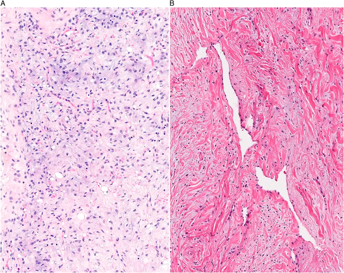
新增了一种肿瘤类型,称为脂肪母细胞瘤样肿瘤,为具有成熟和未成熟脂肪细胞分化的良性梭形细胞肿瘤,同时具有梭形细胞脂肪瘤、黏液样脂肪肉瘤和脂肪母细胞瘤的形态学特征(表1和表2)。3–6其呈分叶状生长,由大小不等的间隔分隔;成熟脂肪细胞、单空泡和/或双空泡脂肪母细胞与温和的梭形细胞混合存在;基质可呈不同程度的黏液样至胶原性改变,并伴有薄壁丛状血管(图1A)。
本版明确了非典型梭形细胞/多形性脂肪瘤样肿瘤的诊断标准,并将其分为非典型梭形细胞和非典型多形性两个亚型。7在高分化脂肪肉瘤和去分化脂肪肉瘤谱系中,无脂肪分化的肿瘤现根据核分裂象计数加以区分:鉴于生存结局的差异,每10个HPF中核分裂象不超过4个的肿瘤归为富于细胞的高分化脂肪肉瘤(非典型脂肪瘤样肿瘤),而核分裂象为5个及以上的肿瘤归为去分化脂肪肉瘤(需注意,这些研究大多未注明HPF直径,因此无法准确确定每mm2的核分裂象数)。8因此,不再推荐使用“低级别去分化脂肪肉瘤”这一名称。黏液样脂肪肉瘤的分级参数得到了更精确的规定,明确区分低级别与较高级别病变。较高级别黏液样脂肪肉瘤定义为:肿瘤中细胞更密集、含圆形细胞的区域超过5%,通常伴黏液样基质减少。
纤维母细胞性和肌纤维母细胞性肿瘤
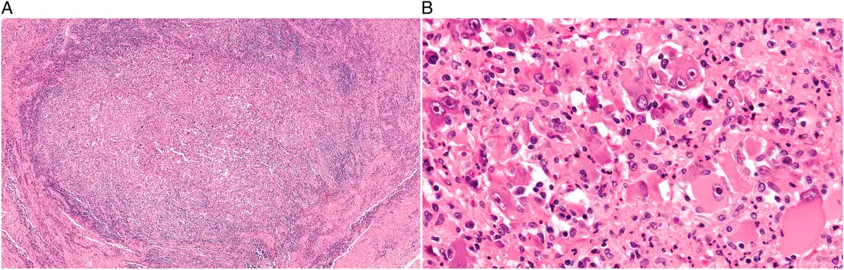
本类别新增了两种肿瘤类型:PRRX1重排的纤维母细胞性肿瘤9,10和结节性坏死性纤维母细胞性肿瘤(见表1和表2)。PRRX1重排的纤维母细胞性肿瘤是一种良性梭形细胞肿瘤,以PRRX1重排为特征,最常见的融合伙伴为NCOA1(图1B)。结节性坏死性纤维母细胞性肿瘤最初被认为是黏液炎性纤维母细胞肉瘤的一个亚型,现已被认定为独立实体,具体定义为一种浅表性间叶性肿瘤,由上皮样细胞构成,伴有显著炎细胞浸润,许多病例可见中央坏死,呈惰性生物学行为(图2)。11这些病变大多具有YAP1::MAML2融合。
命名方面有一些小幅调整:EWSR1::SMAD3阳性的纤维母细胞性肿瘤更名为SMAD3重排的纤维母细胞性肿瘤(预计未来会发现涉及FET基因家族其他成员的融合伙伴);浅表性CD34阳性多形性纤维母细胞性肿瘤的名称中增加了“多形性”一词(因为细胞学多形性对于识别这一肿瘤类型非常重要,同时也可能导致其与多形性肉瘤混淆)。2020年分类中无意遗漏的钙化性纤维性肿瘤已纳入STB6。形态类似硬化性上皮样纤维肉瘤、MUC4阴性且存在涉及YAP1和KMT2A的基因融合的肿瘤,与硬化性上皮样纤维肉瘤归为同一类,同时也认识到,随着更多结局数据的积累,这一归类可能发生变化。12–14值得注意的是,成人型纤维肉瘤已从分类中删除,因为过去诊断为成人型纤维肉瘤的肿瘤,如今基本上都可通过现代方法重新归为其他肉瘤类型(例如单相型滑膜肉瘤、恶性外周神经鞘瘤和隆突性皮肤纤维肉瘤的纤维肉瘤样变异型)。然而,婴儿型纤维肉瘤(IFSC)仍保留在分类中,因为它是发生于儿童、具有特定形态学和遗传学异常的独特肿瘤类型;当IFSC发生于肾脏时,最常用且可接受的名称为“细胞型先天性中胚层肾瘤”。最后,未分化多形性肉瘤(UPS)已从分化未定的肿瘤(此前作为“未分化软组织肉瘤”这一统称下的一个亚型)移至纤维母细胞性和肌纤维母细胞性肿瘤章节,因为尽管称为“未分化”,但在许多病例中,肿瘤细胞似乎显示纤维母细胞分化,有时显示肌纤维母细胞分化。UPS仍被视为排除性诊断。低级别、细胞均一且形态单一的梭形细胞肉瘤不应归为UPS,因为这类肿瘤似乎正越来越多地被归入其他实体。
纤维组织细胞性和滑膜肿瘤
本章在第5版中的标题为“纤维组织细胞性肿瘤”,在STB6中改为“纤维组织细胞性和滑膜肿瘤”。
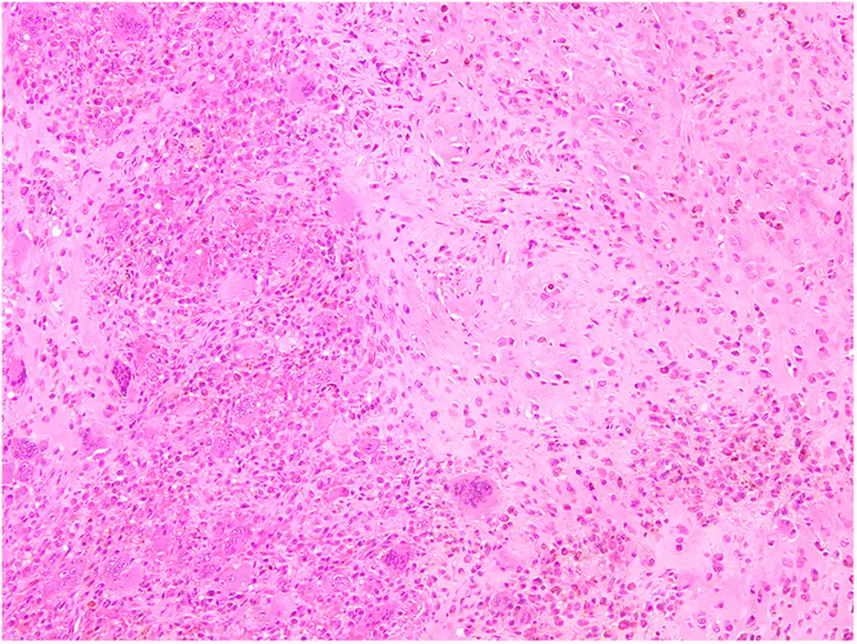
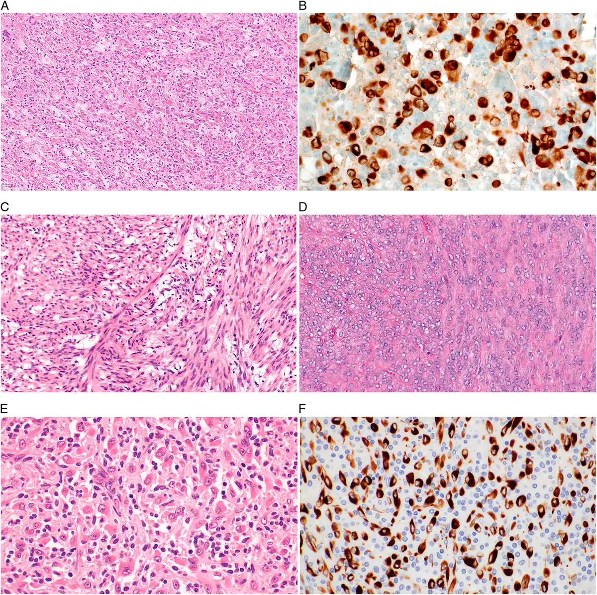
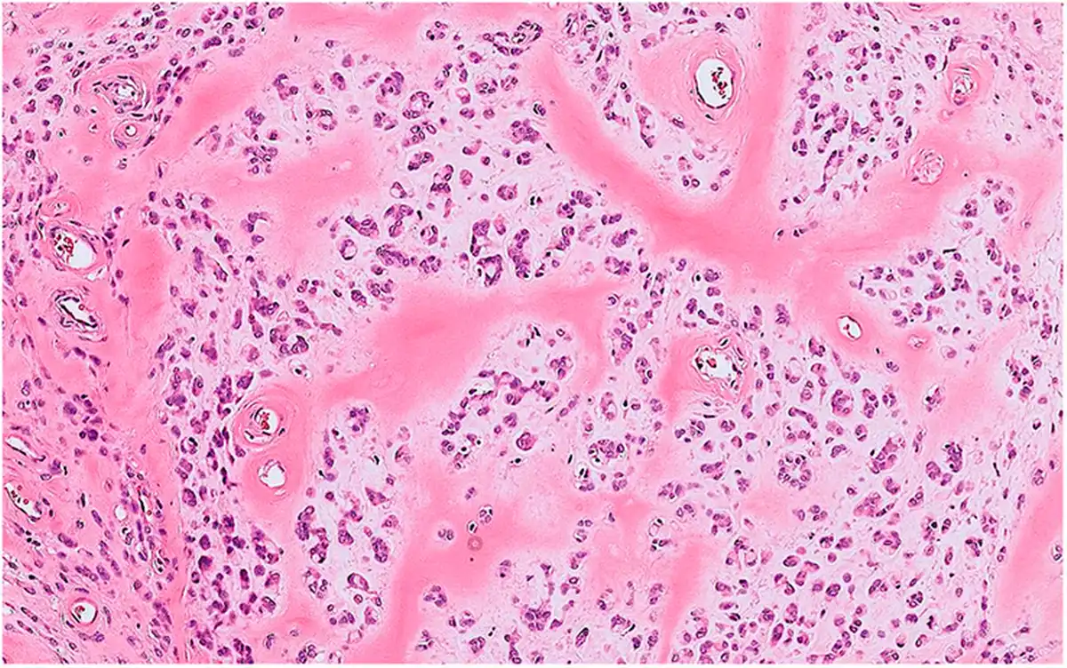
本类别新增了两种肿瘤类型:软骨样滑膜细胞肿瘤和角蛋白阳性富于巨细胞的肿瘤(KPGCT)(见表1和表2)。软骨样滑膜细胞肿瘤(又称“软骨样腱鞘巨细胞瘤”或“钙化性软骨样间叶性肿瘤”)好发于颞下颌关节区域,由不同比例的大型嗜酸性滑膜细胞、破骨细胞样巨细胞、透明软骨和钙化基质构成(图3),常具有FN1重排。15–19KPGCT是一种谱系未定的间叶性肿瘤,由多核巨细胞、混合性炎细胞、组织细胞及少量角蛋白阳性的单核细胞构成(图4A、B),具有HMGA2::NCOR2融合。20–23深部纤维组织细胞瘤已从本分类中删除,因为它属于皮肤良性纤维组织细胞瘤(皮肤纤维瘤)的一个亚型,已纳入WHO皮肤肿瘤分类。
周细胞性(血管周围)肿瘤
新增了一种肿瘤类型,即SRF重排的肌样肿瘤(见表1和表2),涵盖一组以良性为主、具有肌样或未成熟平滑肌分化的肿瘤,其特征为SRF基因与多种伙伴发生融合(图4C、D)。24–26在此前的第5版中,这些肿瘤中的一部分被描述为肌纤维瘤的“非典型富于细胞变异型”。
平滑肌肿瘤
炎性平滑肌肉瘤已从本节删除,因为这一类别的肿瘤现称为炎性横纹肌母细胞性肿瘤(见骨骼肌肿瘤一节)。
骨骼肌肿瘤
本卷纳入了炎性横纹肌母细胞性肿瘤(见表1和表2),其中包括既往称为炎性平滑肌肉瘤的肿瘤。其定义为中间性(罕见转移)肿瘤,特征包括骨骼肌分化、显著的淋巴细胞和组织细胞浸润(图4E、F),以及近单倍体化。27–30可见恶性进展为横纹肌肉瘤,表现为核分裂活性高,且常伴坏死。31根据遗传学特征及临床行为的差异,梭形细胞/硬化性横纹肌肉瘤已分为3个独立实体:融合相关梭形细胞横纹肌肉瘤和MYOD1突变型梭形细胞/硬化性横纹肌肉瘤在软组织章中讨论,而TFCP2重排的横纹肌肉瘤主要发生于骨,因此在骨肿瘤章中讨论(见表1和表2)。融合相关梭形细胞横纹肌肉瘤是一组异质性肉瘤,具有骨骼肌分化及束状排列的梭形细胞形态,以反复出现的特征性融合为定义依据,其融合伙伴包括NCOA2或CITED2、ZFP64和VGLL3等。32–34MYOD1突变型梭形细胞/硬化性横纹肌肉瘤是横纹肌肉瘤的一种侵袭性亚型,以束状排列的梭形细胞和/或硬化性形态及MYOD1 p.L122R突变为特征。31,35–37外胚间叶瘤是一种极罕见且主要发生于儿童的肿瘤,已不再作为独立实体纳入分类;根据其中混杂的神经母细胞性成分,该病变现被认定为胚胎性横纹肌肉瘤的一个亚型,称为“伴分歧性神经外胚层分化的胚胎性横纹肌肉瘤”。
胃肠道间质瘤
鉴于独特的分子发病机制、可能与家族性综合征相关的意义,以及临床行为上的差异,琥珀酸脱氢酶缺陷型胃肠道间质瘤已被认定为独立类别。38–42
软骨和骨性肿瘤
本节未作重大修改。
外周神经鞘肿瘤
上皮样恶性外周神经鞘瘤(EMPNST)此前被视为MPNST的一个亚型,现根据其独特的上皮样形态及特定的分子发病机制(SMARCB1 [INI1]表达缺失)被认定为独立实体。43,44
分化未定的肿瘤
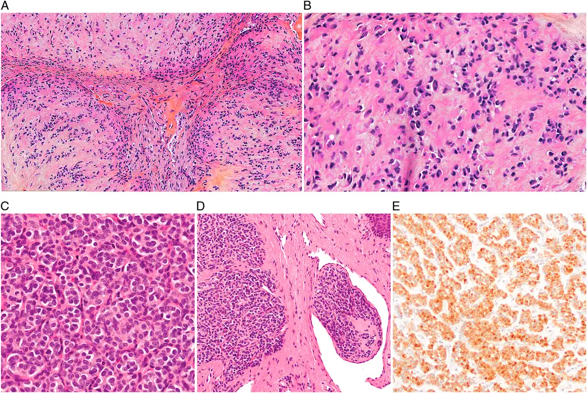
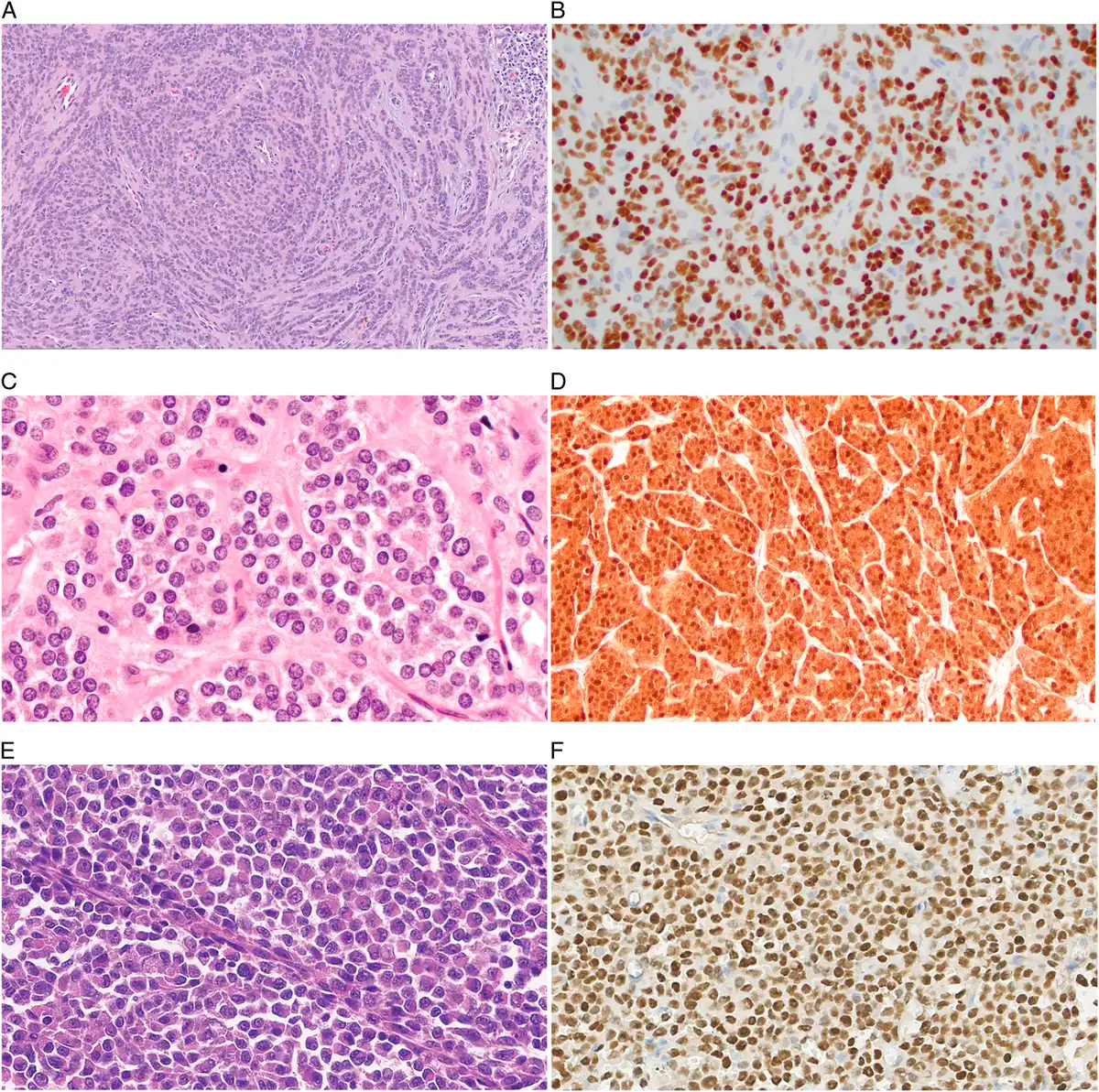
本卷首次纳入了大量近期确立的肿瘤类型,包括OGT重排的间叶性肿瘤(既往称为OGT重排的肢端间叶性肿瘤)(图5)、45肢端纤维软骨黏液样肿瘤(AFCMT)、46GLI1改变的间叶性肿瘤、47–49浅表性FET-ETS家族神经嵴肿瘤、50,51假内分泌肉瘤、52,53NUT-重排肉瘤、54,55以及FET-CREB家族上皮样间叶性肿瘤(见表1和表2)。56–58AFCMT是一种发生于肢端、分化未定的良性间叶性肿瘤,由温和的梭形或圆形细胞构成,细胞分布于软骨黏液样间质内,纤维血管间隔将其分隔成小叶(图6A、B)。GLI1改变的间叶性肿瘤临床行为不一,常呈巢状排列的上皮样形态,并具有丰富的毛细血管网(图6C-E),由GLI1融合或扩增驱动。浅表性FET-ETS家族神经嵴肿瘤是一种惰性浅表肿瘤,具有与尤文肉瘤相似的融合事件(图7A、B)。假内分泌肉瘤是一种谱系未定的恶性肿瘤,主要发生于椎旁软组织,形态类似高分化神经内分泌肿瘤(图7C、D),但不表达角蛋白和神经内分泌标志物,并具有CTNNB1突变。NUT-重排肉瘤是一种侵袭性恶性间叶性肿瘤,以异质性形态(图7E、F)及涉及NUTM1的基因融合为特征。FET-CREB家族恶性上皮样间叶性肿瘤通常累及间皮衬覆的体腔,呈上皮样形态并表达上皮标志物(图8),其特征为EWSR1或FUS基因与CREB转录因子家族成员(ATF1, CREB1和CREM)发生融合。
既往属于“新兴”类别的NTRK重排的梭形细胞肿瘤已扩展为激酶改变的梭形细胞肿瘤,因为目前已认识到,这一肿瘤类别中可检出多种激酶基因融合(以及点突变)。59–61PATZ1重排的肉瘤被纳入本节;该肿瘤在第5版中被归入伴EWSR1非ETS融合的圆细胞肉瘤。这些肿瘤比圆细胞肉瘤具有更大的形态学异质性,可见圆形和梭形细胞形态,共表达肌上皮和骨骼肌标志物,并以涉及EWSR1和PATZ1的融合为特征。两个实体(非典型纤维黄色瘤和异位性错构瘤性胸腺瘤)不再纳入本节;也不再推荐以“未分化软组织肉瘤”这一统称作为杂类,将未分化多形性肉瘤(已移至纤维母细胞性和肌纤维母细胞性肿瘤,见上文)、未分化梭形细胞肉瘤和未分化圆细胞肉瘤等肿瘤归于其中。
软组织和骨的血管肿瘤
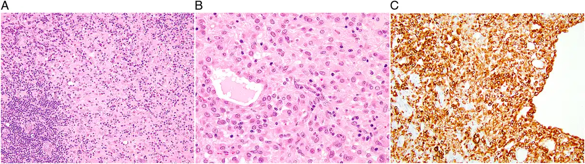
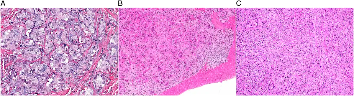
本节最重要的变化是对既往称为血管瘤和淋巴管瘤的病变进行命名调整,引入血管异常(及淋巴管畸形)这一术语,使分类与国际血管异常研究学会(ISSVA)保持一致。62梭形细胞血管瘤在2020年分类中被无意遗漏,现已纳入STB6。鉴于组织病理学、分子发病机制和临床行为方面的差异,YAP1::TFE3血管内皮瘤已从上皮样血管内皮瘤中分离出来(见表1和表2)。63YAP1::TFE3血管内皮瘤是一种恶性血管肿瘤,由上皮样内皮细胞构成,细胞常具有丰富的嗜酸性胞质和血管形成特征(图9A),以YAP1::TFE3融合为特征。该肿瘤类型患者的临床结局优于常规型上皮样血管内皮瘤患者。
未分化梭形细胞和小圆细胞肉瘤
鉴于这些高级别肉瘤中的部分肿瘤不仅具有圆形细胞形态,还具有梭形细胞形态,本章名称作了相应修改。本节纳入的肿瘤包括尤文肉瘤、NFATC2重排的肉瘤、伴BCOR遗传学改变的肉瘤及CIC重排的肉瘤。如前所述,第5版中伴EWSR1非ETS融合的圆细胞肉瘤这一类别已拆分为NFATC2重排的肉瘤(纳入本节)64–66和PATZ1重排的肉瘤(纳入软组织肿瘤章中的分化未定的肿瘤)。NFATC2重排的肉瘤是一种具有纤维玻璃样间质的圆形和梭形细胞肉瘤,伴有EWSR1::NFATC2或FUS::NFATC2融合。
骨肿瘤
骨肿瘤分类的重要变化
软骨源性肿瘤
本版强调,应结合影像学,采用多学科方法诊断这些病变。在第5版中,“非典型软骨性肿瘤”和“1级软骨肉瘤”这两个术语得到了进一步界定:四肢骨骼(长管状骨和短管状骨)的软骨性肿瘤诊断为非典型软骨性肿瘤,而“1级软骨肉瘤”这一术语则专用于中轴骨骼的肿瘤,包括骨盆、肩胛骨和颅底(扁骨),以反映这些部位肿瘤较差的临床结局。67对组织形态相同的肿瘤使用不同术语,可能被认为有悖直觉且存在争议。然而,这种命名既便于应用,又能反映这些部位的肿瘤结局较差、需要更广泛手术切除的情况,具有双重益处。67因此,现行分类进一步认可了这一命名。基于软骨成分的存在,胸壁软骨间叶性错构瘤在STB6中归入软骨源性肿瘤进行讨论,而非像STB5中那样归入其他骨肿瘤。根据近期发表的文献,68STB6现已认识到,极少数软骨母细胞瘤为恶性。这些肿瘤具有异型性和渗透性生长,发生于年龄较大者的扁骨,IHC显示H3K36M阳性。
成骨性肿瘤
良性成骨性肿瘤包括骨瘤、骨样骨瘤和骨母细胞瘤。骨肉瘤是最常见的恶性骨肉瘤,根据部位及形态可区分多种类型和形态学模式。与第5版相比,其分类未作修改。
纤维源性肿瘤
“纤维源性肿瘤”这一类别已从骨肿瘤分类中删除。骨的促结缔组织增生性纤维瘤移至“其他骨肿瘤”。骨纤维肉瘤过去被视为排除性诊断,现已删除(与软组织章相同),因为其中大多数似乎是去分化软骨肉瘤或骨平滑肌肉瘤,或是软组织肿瘤罕见的骨内表现,例如孤立性纤维性肿瘤、激酶改变的梭形细胞肿瘤或滑膜肉瘤。
富于破骨细胞样巨细胞的肿瘤
虽然许多骨肿瘤可含有破骨细胞,但在骨巨细胞瘤、动脉瘤样骨囊肿和非骨化性纤维瘤中,破骨细胞是其定义性特征。研究发现,除甲状旁腺激素(PTH)水平升高外,KRAS突变也是甲状旁腺功能亢进性棕色瘤发生所必需的条件,从而证实其具有肿瘤性;69STB6将其纳入为独立实体。其定义为发生于甲状旁腺功能亢进背景下、混有破骨细胞样巨细胞的良性梭形细胞肿瘤(图9B)。
上一版已提及,不伴巨细胞瘤组织学表现的骨的肉瘤(骨肉瘤、未分化多形性肉瘤)中也可出现H3.3 p.G35W (G34W),提示其与骨巨细胞瘤存在关联。较新的证据表明,具有H3.3 p.G35 (G34)突变的未分化多形性肉瘤或骨肉瘤显示相似的分子和甲基化谱,因此可能代表恶性骨巨细胞瘤。70然而,仍需更大队列的研究来证实这一关联并指导临床管理。
脊索源性肿瘤
脊索源性肿瘤包括良性脊索细胞瘤、常规型脊索瘤(包括软骨样脊索瘤)、去分化脊索瘤和低分化脊索瘤。与第5版相比,其分类未作修改。
其他骨肿瘤
在第6版中,“骨纤维结构不良(OFD)样造釉细胞瘤”更名为“非典型OFD”,现被视为OFD的一种局部侵袭性亚型。这更能反映非典型OFD的非恶性行为,并应有助于避免对年轻患者的这类肿瘤进行过度治疗。71不过,诊断仍可能困难,观察者间差异较大。TFCP2重排的横纹肌肉瘤是一种具有骨骼肌分化的新型高级别肉瘤(图9C),以TFCP2与EWSR1或FUS融合为特征,好发于颌面骨。72该肿瘤现作为新的骨肿瘤实体纳入分类(见表1和表2)。
软组织和骨的造血及树突状细胞肿瘤
造血及树突状细胞肿瘤的重要变化
STB6仅描述发生于非黏膜的结外软组织或原发累及骨的造血系统肿瘤,按照这一原则,本章纳入浆细胞瘤、累及骨的弥漫性大B细胞淋巴瘤非特指型(NOS)、朗格汉斯细胞组织细胞增生症、Erdheim-Chester病、滤泡树突状细胞肉瘤及Rosai-Dorfman病。
发生于骨髓腔内的髓系肿瘤及其他骨髓内增生性病变不纳入本章。但还应注意,在局部广泛病变或系统性疾病的情况下,几乎任何造血淋巴系统肿瘤均可继发累及软组织和/或骨。因此,诊断评估需整合形态学、免疫表型和分子检测结果,并结合适当的影像学及临床评估,以确定受累范围,并判断病变是否为软组织或骨的原发性肿瘤。
软组织和骨的遗传性肿瘤综合征
遗传性肿瘤综合征的重要变化
| 疾病/表型 | MIM号* | 遗传方式 | 位点 | 基因 | 基因/位点MIM号 | 蛋白 | 蛋白的正常功能 |
|---|---|---|---|---|---|---|---|
| 内生软骨瘤病(Ollier病和Maffucci综合征) | 166000 | PZ | 2q34 | IDH1 | 147700 | IDH1 | 异柠檬酸脱氢酶;将异柠檬酸转化为α-酮戊二酸;产生NADPH并调控线粒体氧化还原平衡 |
| 15q26.1 | IDH2 | 147650 | IDH2 | ||||
| Li-Fraumeni综合征 | 151623 | AD | 17p13.1 | TP53 | 191170 | TP53 (p53) | DNA损伤应答,“基因组守护者” |
| McCune-Albright综合征 | 174800 | PZ | 20q13.32 | GNAS | 139320 | Gsα | G蛋白的α亚基;将信号从7次跨膜受体传递至第二信使cAMP |
| 多发性骨软骨瘤 | 133700 | AD | 8q24.11 | EXT1 | 608177 | EXT1 | EXT1和EXT2形成异源寡聚复合物,参与硫酸乙酰肝素的生成;对骨和软骨形成至关重要 |
| 133701 | AD | 11p11.2 | EXT2 | 608210 | EXT2 | ||
| 毛发-鼻-指(趾)骨综合征2型 | 150230 | 8q24.q11-q24.13 | 涉及EXT1的连续基因缺失综合征 | ||||
| Potocki-Shaffer综合征 | 601224 | 11p11.2 | 涉及EXT2的连续基因缺失综合征 | ||||
| 1型神经纤维瘤病 | 162200 | AD | 17q11.2 | NF1 | 613113 | 神经纤维瘤蛋白(NF1) | 调控RAS信号传导 |
| 骨Paget病 | — | AD | 17p13.2 | PFN1 | 176610 | PFN1 | 调控NFκB信号传导 |
| 167250 | AD | 5q35.3 | SQSTM1 | 601530 | SQSTM1 (p62) | 调控NFκB信号传导 | |
| 602080 | AD | 18q21.33 | TNFRSF11A | 603499 | RANK | 破骨细胞的主要调节因子 | |
| 239000 | AR | 8q24.12 | TNFRSF11B | 602643 | 骨保护素 | TNFSF11/RANKL的诱饵受体 | |
| 616833 | AD | 1q21.3 | ZNF687 | 610568 | ZNF687 | 未知 |
AD表示常染色体显性遗传;AR表示常染色体隐性遗传;PZ表示合子后。
* OMIM表示在线人类孟德尔遗传数据库(https://www.omim.org)。
通过应用大规模平行DNA测序等测序技术,在越来越多的基因中发现了与软组织和/或骨肿瘤发生风险升高相关的胚系(体质性)致病变异(表3)。这些肿瘤易感性遗传变异常伴有其他表型影响,形成临床上可识别的综合征。由于胚系致病变异常遗传自父母一方,识别这类肿瘤易感综合征十分重要,有助于在受累家系中筛查高风险个体,转诊进行遗传咨询,并按指南实施监测,以早期发现癌症。虽然这些综合征大多数呈孟德尔遗传,但目前也已识别出由合子后突变导致疾病的患者,他们可能需要不同的治疗方式及个体化随访。对胚系肿瘤易感变异的研究可能有助于阐明散发性肿瘤的发病机制。
本章详细介绍了6种与骨或软组织肿瘤发生易感性相关的综合征(见表3)。之所以纳入这些特征明确的综合征,是因为骨或软组织肿瘤作为综合征的一部分出现频率较高,且是导致该综合征的遗传学改变的主要后果,其肿瘤特征往往与相应的散发性肿瘤不同。
STB6中与遗传性肿瘤综合征有关的变化包括:Rothmund-Thomson综合征和Werner综合征不再像第5版那样各设独立章节,而仅在引言中的一张表内提及。骨Paget病则由第5版引言表格中的条目调整为本版中的一个详细章节。
结论
第6版软组织和骨肿瘤卷基于目前可获得的最佳证据,为发生于软组织和骨的肿瘤提供了更新的分类和全面概述。本版力求在兼顾科学进展的同时满足全球需求。
致谢
作者衷心感谢WHO肿瘤分类(WCT)工作组全体成员提供的支持,以及WCT各分委员会所作的贡献,包括遗传学与基因组学、计算病理学、WCT/中低收入国家(LMIC)联络、肿瘤分类协调、影像学与放射学,以及肿瘤定义与澄清分委员会。图片经许可转载自:WHO肿瘤分类编委会。《软组织和骨肿瘤》。里昂(法国):国际癌症研究机构;2026年。(WHO肿瘤分类丛书,第6版,第3卷)。获取地址:https://tumourclassification.iarc.who.int/chapters/87。
- STB6 includes soft tissue tumors (n=116), bone tumors (n=46), vascular tumors of soft tissue and bone (n=18), undifferentiated spindle and small round cell sarcomas (n=4), hematopoietic and dendritic cell neoplasms (n=6) and genetic syndromes (n=6) — 62 sarcomas in total; 19 new entities were added.
- Inclusion threshold for new entities: ≥5 published cases from 2 independent groups in peer-reviewed journals; borderline entities are “provisional” rather than “emerging”; “variant” is replaced by “morphological pattern”.
- “Essential” criteria are mostly morphology plus basic IHC (molecular confirmation essential for molecularly defined tumors such as BCOR-altered sarcomas); “desirable” criteria add molecular tests and surrogate IHC; ICD-O-4 morphology codes are given for every type and subtype.
- Tables 1–2 summarise new and deleted entities (e.g. new PRRX1-rearranged fibroblastic tumor, keratin-positive giant cell-rich tumor, SRF-rearranged myoid neoplasm, YAP1::TFE3 hemangioendothelioma, brown tumor of hyperparathyroidism; deleted adult fibrosarcoma, deep fibrous histiocytoma, inflammatory leiomyosarcoma, ectomesenchymoma, fibrosarcoma of bone); Table 3 covers genetic tumor syndromes; 9 figure plates.
Scope: open-access full text (CC BY-NC-ND 4.0) from the AJSP publisher page, including 9 figures, 3 tables (with notes) and acknowledgments; the reference list and the WCT Standing Editorial Board roster are not included. Superscript numbers are the original reference numbers.
The World Health Organization (WHO) classification of Tumors (WCT) provides a uniform nomenclature and standardized framework for categorizing tumors of soft tissue and bone, facilitating accurate diagnosis, guiding treatment, informing research, epidemiological reporting, and the development of international reporting and treatment guidelines. The 6th edition of the WHO Classification of soft tissue and bone tumors (STB6) is an update of the 5th edition classification, encompassing some major and minor changes. The 6th edition integrates the latest scientific and molecular discoveries with decades of pathological expertise to establish a globally accepted common language for soft tissue and bone tumor diagnosis and management. The criteria used for considering inclusion of new entities into this volume included publication of at least 5 cases of the neoplastic entity by 2 independent groups in peer-reviewed journals; any borderline entities included are referred to as “provisional” rather than “emerging.”
In this volume, the “essential and desirable diagnostic criteria” for tumors have been developed considering different resource settings, ensuring diagnostic accuracy while maintaining flexibility. The “essential criteria” are mostly morphology-based, including certain essential, invariably “basic” immunohistochemical (IHC) stains, except for molecularly defined tumors such as sarcomas with BCOR alterations, for which molecular confirmation is considered essential. The “desirable criteria” include an additional diagnostic layer of molecular tests and/or “newer” resources, including surrogate IHC markers. The morphology codes of the updated International Classification of Diseases Oncology, 4th iteration (ICDO-4) have been included for each tumor type and subtype in STB6, ensuring and facilitating more accurate and granular epidemiological data collection.1 Inclusion of tumor types and subtypes is based on better definitions. A tumor type (entity) is a tumor in which multiple parameters (eg, clinical, anatomic, histopathological, and/or molecular features) differ from those of other tumor types. A subtype is recognized as being different in at least one dimension from the main tumor type (clinical, histopathological, or genetic), preferably resulting in a different treatment or outcome. A morphological pattern (no longer called “variant”) is defined as a specific morphology of a given tumor type, important for pathologists to be aware of to facilitate recognition, but not associated with different clinical behavior.
The classification was drafted by 203 editors and authors from 27 countries, representing radiology, medical oncology, genetics, surgical oncology, and orthopedic surgery, in addition to pathology. When appropriate, responsible authors from the previous 5th edition were acknowledged for their contributions, providing the basis for the 6th edition. A consensus was subsequently reached among the expert editorial board. Entities with specific organ-restricted presentation, such as biphenotypic sinonasal sarcoma or uterine sarcomas, are excluded from STB6 and instead discussed in other WCT volumes, with the exception of gastrointestinal stromal tumors, as in many centers, these are treated by sarcoma surgeons and medical oncologists and are discussed in sarcoma multidisciplinary teams.
CHANGES IN HISTONE GENE ANNOTATIONS
With regard to the annotation of histone H3 protein nomenclature, a hybrid format is used as a practical compromise that preserves the widely recognized legacy terminology (which uses protein numbering excluding the initiating methionine that is cleaved from the mature protein) while remaining aligned with genetic reporting standards;2 specifically, the 3-letter amino acid description is omitted and the legacy terminology is given in parentheses, for example, H3.3 p.G35W (G34W) in giant cell tumor of bone. For IHC markers targeting histone proteins, we retain the established epitope-based terminology (eg, H3K36M and H3K27me3) without the “p.” prefix and with the conventionally used formatting (eg, H3K27me3 without spaces). These terms refer to protein expression or posttranslational modifications rather than genomic alterations and are therefore most consistent with current diagnostic practice and the published literature.
CHANGES IN THE CLASSIFICATION FRAMEWORK OF STB6
In the 5th edition, separate sections on vascular tumors were included in both the soft tissue and bone tumor chapters, which was mainly driven by their different clinical behavior. Vascular tumors in bone often present as multifocal disease, and epithelioid hemangioma is locally aggressive in bone, while benign in soft tissue. In this edition, these sections were combined to limit redundancy; differences between bone and soft tissue presentation are clearly marked within the respective sections.
STB6 has also taken a different approach to hematolymphoid tumors of the soft tissues and bone, combining them into one chapter. Tumors included in this chapter are hematopoietic neoplasms typically derived from lymphoid, plasma cell, or histiocytic/dendritic cell lineages that arise in nonmucosal extranodal soft tissues or involve bone primarily, often presenting as mass-forming and/or destructive osseous lesions.
In other WHO 6th edition books (released and upcoming), various soft tissue tumor entities are presented in a harmonized way, mostly within a combined chapter of “mesenchymal tumors.”
SOFT TISSUE TUMORS
Significant Changes in Soft Tissue Tumor Classification
| Chapter | Newly added tumors | Deleted tumors |
|---|---|---|
| Soft tissue tumors | ||
| Adipocytic tumors | Lipoblastoma-like tumor | |
| Fibroblastic and myofibroblastic tumors | PRRX1-rearranged fibroblastic tumor Nodular necrotizing fibroblastic tumor Calcifying fibrous tumor* | Adult fibrosarcoma |
| Fibrohistiocytic and synovial tumors | Chondroid synoviocytic neoplasm Keratin-positive giant cell-rich tumor | Deep fibrous histiocytoma |
| Pericytic (perivascular) tumors | SRF-rearranged myoid neoplasm | |
| Smooth muscle tumors | Inflammatory leiomyosarcoma | |
| Skeletal muscle tumors | Inflammatory rhabdomyoblastic tumor Fusion-associated spindle cell rhabdomyosarcoma† MYOD1-mutant spindle cell/sclerosing rhabdomyosarcoma† | Ectomesenchymoma |
| Gastrointestinal stromal tumor | Succinate dehydrogenase-deficient gastrointestinal stromal tumor | |
| Tumors of uncertain differentiation | OGT-rearranged mesenchymal neoplasm (formerly known as OGT-rearranged acral mesenchymal neoplasm) Acral fibrochondromyxoid tumor GLI1-altered mesenchymal tumors Superficial FET-ETS family neurocristic tumor Pseudoendocrine sarcoma NUT-rearranged sarcoma FET-CREB family epithelioid mesenchymal tumor PATZ1-rearranged sarcoma‡ | |
| Vascular tumors of soft tissue and bone | YAP1::TFE3 hemangioendothelioma Spindle cell hemangioma* | |
| Undifferentiated spindle and small round cell sarcomas | NFATC2-rearranged sarcoma‡ | |
| Bone tumors | ||
| Osteoclastic giant cell-rich tumors | Brown tumor of hyperparathyroidism | |
| Fibrogenic tumors§ | Fibrosarcoma of bone | |
| Other tumors of bone | TFCP2-rearranged rhabdomyosarcoma† |
* Inadvertently omitted in the 2020 WCT.
† Spindle cell/sclerosing rhabdomyosarcoma has been split into these 3 separate entries.
‡ The category round cell sarcomas with EWSR1-non-ETS fusions has been split into NFATC2-rearranged sarcoma and PATZ1-rearranged sarcoma.
§ The family of fibrogenic tumors that was listed under bone tumors in STB5 has been removed in STB6.
| Entity | Behavior | Section | Key molecular | Morphology | Note |
|---|---|---|---|---|---|
| Lipoblastoma-like tumor | Benign | Adipocytic | No fusions | Reminiscent of lipoblastoma and myxoid liposarcoma | |
| PRRX1-rearranged fibroblastic tumor | Benign | Fibroblastic/myofibroblastic | PRRX1 rearrangement | Paucicellular circumscribed bland spindle cell neoplasm; hyalinized stroma with variably wire-like and ropey collagen; prominent ectatic and irregular vascular spaces | |
| Nodular necrotizing fibroblastic tumor | Intermediate | Fibroblastic/myofibroblastic | YAP1::MAML2 fusion | Well-defined nodules of neoplastic cells with surrounding and/or admixed inflammation; tumor cells with copious, eosinophilic-to-amphophilic cytoplasm and enlarged, vesicular nuclei with prominent nucleoli; often central necrosis. | |
| Chondroid synoviocytic neoplasm | Intermediate | Fibrohistiocytic/synovial | FN1 fusions (various partners) | Nodules of large, eosinophilic, moderately pleomorphic synoviocytes, surrounded by calcified matrix and cartilage, with sparse histiocytes, osteoclasts, and foamy macrophages. | Temporomandibular joint region |
| Keratin-positive giant cell-rich tumor | Intermediate | Fibrohistiocytic/synovial | HMGA2::NCOR2 | Polymorphous population of giant cells (osteoclast-type or Touton), histiocytes, inflammatory cells, and keratin-positive mononuclear cells | |
| SRF-rearranged myoid neoplasm | Benign | Pericytic (perivascular) | SRF gene fusions (various partners) | Monomorphic ovoid to spindle cells with fibrillary cytoplasm; arranged in short fascicles; myoid or immature smooth muscle differentiation | |
| Inflammatory rhabdomyoblastic tumor | Intermediate | Skeletal muscle | Near haploidy | Spindled or epithelioid cells with eosinophilic cytoplasm, variable degrees of nuclear atypia, and low mitotic activity; massive intratumoral lymphohistiocytic infiltration; skeletal muscle differentiation | Previously “inflammatory leiomyosarcoma” |
| Fusion-associated spindle cell rhabdomyosarcomas | Malignant | Skeletal muscle | Fusions due to rearrangement of genes in various combinations (ZFP64, NCOA2, NCOA3, VGLL3, CITED2, and others) | Monomorphic spindle cell morphology; expression of at least 2 skeletal muscle markers | Split from spindle cell/sclerosing RMS; variable prognosis depending on fusions |
| MYOD1-mutant spindle cell/sclerosing rhabdomyosarcoma | Malignant | Skeletal muscle | MYOD1 mutation p.L122R | Spindle cells arranged in fascicles and/or oval to spindle cells with variable amount of sclerotic stroma; MYOD1 typically diffuse and intense | Split from spindle cell/sclerosing RMS; aggressive; poor prognosis |
| OGT-rearranged mesenchymal neoplasm | Benign | Uncertain differentiation | OGT rearrangement | Cords or nests of epithelioid cells within a hyalinized or fibromyxoid stroma | Acral location |
| Acral fibrochondromyxoid tumor | Benign | Uncertain differentiation | THBS1 rearrangement (frequently with ADGRF5 or rarely other) | Multinodular tumor with fibrous, myxoid or chondroid stroma; fibrovascular septa; bland spindle or round cells; CD34 and ERG positive | Acral location |
| GLI1-altered mesenchymal tumors | Variable | Uncertain differentiation | GLI1 amplification or rearrangement | Lobular architecture; nested growth; epithelioid or ovoid cytology; prominent admixed branching capillary vessels | |
| Superficial FET-ETS family neurocristic tumor | Benign | Uncertain differentiation | FET-ETS family fusions (EWSR1 or FUS and FLI1, ERG, ETV1, ETV5, and others) | Nests, cords, and trabeculae of small, bland, round to ovoid cells; lack of significant cytologic atypia or tumor necrosis; strong and diffuse expression of SOX10, S100 protein, and CD99 | |
| Pseudoendocrine sarcoma | Malignant | Uncertain differentiation | CTNNB1 alteration | Mimics neuroendocrine tumor morphologically; lacks expression of keratins and neuroendocrine markers; β-catenin nuclear positivity | Paravertebral soft tissues |
| NUT-rearranged sarcoma | Malignant | Uncertain differentiation | NUT family (mainly NUTM1) rearrangement (various partners) | Heterogeneous: pure or mixed population of round-epithelioid-spindle cells with monomorphic nuclei; arranged in sheets, nests or fascicles; variable collagenous-myxoid stroma, NUT positive | Highly aggressive |
| FET-CREB family epithelioid mesenchymal tumor | Malignant | Uncertain differentiation | FET-CREB family fusions (EWSR1 or FUS and CREM, ATF1, or CREB1) | Monomorphic epithelioid to round cells; keratin positive | Mesothelial-lined cavities |
| YAP1::TFE3 hemangioendothelioma | Malignant | Vascular tumors | YAP1::TFE3 fusion | Solid growth and/or well-formed vascular channels lined by epithelioid endothelial cells with abundant eosinophilic cytoplasm; TFE3 positive | Now separated from epithelioid hemangioendothelioma |
| Brown tumor of hyperparathyroidism | Benign | Osteoclastic giant cell-rich | KRAS mutation | Monomorphic spindle cells in a storiform pattern; osteoclastic giant cells; hemorrhage | Elevated PTH; regresses with PTH normalization |
| TFCP2-rearranged rhabdomyosarcoma | Malignant | Other bone tumors | TFCP2 rearrangement (EWSR1::TFCP2 or FUS::TFCP2) | Epithelioid, spindle cell, or mixed appearance; skeletal muscle differentiation; co-expression of keratins and ALK | Predilection for maxillofacial bones; highly aggressive |
Adipocytic Tumors
One new tumor type is introduced as lipoblastoma-like tumor, a benign spindle cell neoplasm of mature and immature adipocytic differentiation sharing morphologic features of spindle cell lipoma, myxoid liposarcoma, and lipoblastoma (Tables 1 and 2).3–6 It has a lobulated growth pattern, delineated by variably sized septa; an admixture of mature adipocytes, univacuolated and/or bivacuolated lipoblasts, and bland spindle cells; with variably myxoid to collagenous matrix and thin-walled plexiform vasculature (Fig. 1A).
The diagnostic criteria for atypical spindle cell/pleomorphic lipomatous tumors are clarified in this edition, and they are now separated into atypical spindle cell and atypical pleomorphic subtypes.7 Nonlipogenic tumors within the spectrum of well-differentiated liposarcoma and dedifferentiated liposarcoma are now distinguished based on mitotic rate: owing to survival differences, tumors with up to 4 mitoses per 10 HPF are classified as cellular well-differentiated liposarcoma (atypical lipomatous tumor), while tumors with 5 mitoses or more are classified as dedifferentiated liposarcoma (of note, HPF diameter was not indicated for most of these studies, so the number of mitoses/mm2 cannot be accurately determined).8 The designation “low-grade dedifferentiated liposarcoma” is therefore no longer recommended. More precise grading parameters for myxoid liposarcoma are specified, with explicit distinction between low-grade and higher-grade disease. Higher-grade myxoid liposarcoma is defined by the presence of more cellular areas with round cells exceeding 5% of the tumor, usually accompanied by diminished myxoid matrix.
Fibroblastic and Myofibroblastic Tumors
Two new tumor types are included under this category as PRRX1-rearranged fibroblastic tumor9,10 and nodular necrotizing fibroblastic tumor (see Tables 1 and 2). PRRX1-rearranged fibroblastic tumor is a benign spindle cell neoplasm characterized by PRRX1 rearrangements, most often with NCOA1 (Fig. 1B). While initially suggested to be a subtype of myxoinflammatory fibroblastic sarcoma, nodular necrotizing fibroblastic tumor is now recognized as a separate entity specified as a superficial mesenchymal neoplasm composed of epithelioid cells with a brisk inflammatory infiltrate and, in many cases, central necrosis, showing indolent behavior (Fig. 2).11 Most of these lesions harbor YAP1::MAML2 fusions.
There were small changes in nomenclature: EWSR1::SMAD3-positive fibroblastic tumor was modified to SMAD3-rearranged fibroblastic tumor (anticipating the future identification of fusion partners involving other members of the FET family of genes), and the word “pleomorphic” was added to superficial CD34-positive pleomorphic fibroblastic tumor (given the importance of cytologic pleomorphism in both recognition of this tumor type and possible confusion with pleomorphic sarcomas). Calcifying fibrous tumor, inadvertently omitted from the 2020 classification, has been included in STB6. Tumors that resemble sclerosing epithelioid fibrosarcoma with MUC4 negativity and the presence of gene fusions involving YAP1 and KMT2A were co-classified with sclerosing epithelioid fibrosarcoma, recognizing that this may change as more outcome data are collected.12–14 Notably, adult fibrosarcoma has been removed from the classification, as essentially all tumors diagnosed as adult fibrosarcoma in the past can now be reclassified as other sarcoma types using contemporary methods (eg, monophasic synovial sarcoma, malignant peripheral nerve sheath tumor, and the fibrosarcomatous variant of dermatofibrosarcoma protuberans). However, infantile fibrosarcoma (IFSC) remains in the classification, as it is a distinctive tumor type occurring in children with specific morphological and genetic abnormalities; when IFSC presents in the kidney, the most common and acceptable terminology is “cellular congenital mesoblastic nephroma.” Finally, undifferentiated pleomorphic sarcoma (UPS) has been moved from tumors of uncertain differentiation (where it was considered a subtype under the umbrella term of “undifferentiated soft tissue sarcoma”) to the section on fibroblastic and myofibroblastic tumors, since in many cases the tumor cells appear to show fibroblastic and sometimes myofibroblastic differentiation, despite the “undifferentiated” designation. UPS is still considered a diagnosis of exclusion. Low-grade, uniform and monomorphic spindle cell sarcomas should not be classified as UPS, as they increasingly seem to be resolving into other entities.
Fibrohistiocytic and Synovial Tumors
This chapter’s heading, “fibrohistiocytic tumors” in the 5th edition, is changed to “Fibrohistiocytic and synovial tumors” in STB6.
Two new tumor types are included here as chondroid synoviocytic neoplasm and keratin-positive giant cell-rich tumor (KPGCT) (seeTables 1 and 2). Chondroid synoviocytic neoplasm (a.k.a. “chondroid tenosynovial giant cell tumor” or “calcified chondroid mesenchymal neoplasm”) has a predilection for the temporomandibular joint region, is composed of varying amounts of large, eosinophilic synoviocytes, osteoclastic giant cells, hyaline cartilage, and calcified matrix (Fig. 3), and frequently harbors FN1 rearrangements.15–19 KPGCT is a mesenchymal neoplasm of uncertain lineage, composed of multinucleated giant cells, mixed inflammatory cells, histiocytes, and a small number of keratin-positive mononuclear cells (Fig. 4A, B), harboring HMGA2::NCOR2 fusions.20–23 Deep fibrous histiocytoma has been removed from this classification, since it is a subtype of cutaneous benign fibrous histiocytoma (dermatofibroma), which is included in the WHO classification of skin tumors.
Pericytic (Perivascular) Tumors
A new tumor type, SRF-rearranged myoid neoplasm, has been introduced (see Tables 1 and 2), encompassing a group of predominantly benign tumors exhibiting myoid or immature smooth muscle differentiation and characterized by SRF gene fusions with various partners (Fig. 4C, D).24–26 In the previous 5th edition, a subset of these tumors was described as an “atypical cellular variant” of myofibroma.
Smooth Muscle Tumors
Inflammatory leiomyosarcoma has been omitted from this section, as tumors in this category are now designated as inflammatory rhabdomyoblastic tumor (see the Skeletal Muscle Tumors section).
Skeletal Muscle Tumors
Inflammatory rhabdomyoblastic tumor is introduced in this volume (see Tables 1 and 2), including tumors formerly designated inflammatory leiomyosarcoma. It is defined as an intermediate (rarely metastasizing) neoplasm, characterized by skeletal muscle differentiation, a marked lymphohistiocytic infiltrate (Fig. 4E, F), and near-haploidization.27–30 Malignant progression to rhabdomyosarcoma can be seen and is characterized by high mitotic activity and often necrosis.31 Based on genetic features and differences in clinical behavior, spindle cell/sclerosing rhabdomyosarcoma has been split into 3 separate entities: fusion-associated spindle cell rhabdomyosarcomas and MYOD1-mutant spindle cell/sclerosing rhabdomyosarcoma are discussed in the soft tissue chapter, while TFCP2-rearranged rhabdomyosarcoma predominantly occurs in bone and is discussed in the bone chapter (see Tables 1 and 2). Fusion-associated spindle cell rhabdomyosarcomas are a heterogeneous group of sarcomas with skeletal muscle differentiation and fascicular spindle cell morphology, defined by recurrent pathognomonic fusions, including, among others, NCOA2 or CITED2, ZFP64, and VGLL3 as fusion partners.32–34MYOD1-mutant spindle cell/sclerosing rhabdomyosarcoma is an aggressive subtype of rhabdomyosarcoma, characterized by fascicular spindle cell and/or sclerosing morphology and MYOD1 p.L122R mutation.31,35–37 Ectomesenchymoma, an extremely rare and predominantly pediatric tumor, was removed as a separate entity from the classification; this lesion is now recognized as a subtype of embryonal rhabdomyosarcoma and referred to as “embryonal rhabdomyosarcoma with divergent neuroectodermal differentiation” based on the presence of intermixed neuroblastic elements.
Gastrointestinal Stromal Tumor
Succinate dehydrogenase-deficient gastrointestinal stromal tumor has been recognized as a separate category, given the distinct molecular pathogenesis, implications for possible familial syndromic associations, and differences in clinical behavior.38–42
Chondro-Osseous Tumors
No significant changes were made to this section.
Peripheral Nerve Sheath Tumors
Epithelioid malignant peripheral nerve sheath tumor (EMPNST), previously considered a subtype of MPNST, is now recognized as a separate entity, based on its distinctive epithelioid morphology and specific molecular pathogenesis (loss of SMARCB1 [INI1] expression).43,44
Tumors of Uncertain Differentiation
A large number of recently recognized tumor types are included for the first time in this volume, including OGT-rearranged mesenchymal neoplasm (formerly known as OGT-rearranged acral mesenchymal neoplasm) (Fig. 5),45 acral fibrochondromyxoid tumor (AFCMT),46GLI1-altered mesenchymal tumors,47–49 superficial FET-ETS family neurocristic tumor,50,51 pseudoendocrine sarcoma,52,53 NUT-rearranged sarcoma,54,55 and FET-CREB family epithelioid mesenchymal tumors (see Tables 1 and 2).56–58 AFCMT is a benign mesenchymal neoplasm of uncertain differentiation arising in acral sites and composed of bland spindle or round cells arranged in a chondromyxoid stroma lobulated by fibrovascular septa (Fig. 6A, B). GLI1-altered mesenchymal tumors have variable clinical behavior, often nested epithelioid morphology and a rich capillary network (Fig. 6C-E), and are driven by fusions or amplification of GLI1. Superficial FET-ETS family neurocristic tumor is an indolent superficial neoplasm that shares similar fusion events with Ewing sarcoma (Fig. 7A, B). Pseudoendocrine sarcoma is a malignant neoplasm of uncertain lineage, predominantly arising in paravertebral soft tissues, morphologically resembling a well-differentiated neuroendocrine tumor (Fig. 7C, D), but lacking expression of keratins and neuroendocrine markers, while harboring CTNNB1 mutations. NUT-rearranged sarcoma is an aggressive malignant mesenchymal neoplasm characterized by heterogeneous morphology (Fig. 7E, F) and gene fusions involving NUTM1. FET-CREB family malignant epithelioid mesenchymal tumors typically involve mesothelial-lined cavities, show epithelioid morphology and epithelial marker expression (Fig. 8), and are characterized by fusions between EWSR1 or FUS genes with members of the CREB family of transcription factors (ATF1, CREB1, and CREM).
The former “emerging” category of NTRK-rearranged spindle cell neoplasms was expanded to kinase-altered spindle cell neoplasms, since it is now recognized that a wide range of kinase gene fusions (as well as point mutations) can be identified in this tumor class.59–61PATZ1-rearranged sarcoma was introduced into this section; this tumor was included within round cell sarcomas with EWSR1-non-ETS fusions in the 5th edition. These tumors show more morphologic heterogeneity than round cell sarcomas, with round and spindle cell morphology, co-express myoepithelial and skeletal muscle markers, and are characterized by fusions involving EWSR1 and PATZ1. Two entities (atypical fibroxanthoma and ectopic hamartomatous thymoma) were no longer included, and the umbrella term “undifferentiated soft tissue sarcoma” is no longer recommended as a miscellaneous category for the group of tumors including undifferentiated pleomorphic sarcoma (moved to (myo)fibroblastic tumors; see above), undifferentiated spindle cell sarcoma, and undifferentiated round cell sarcoma.
VASCULAR TUMORS OF SOFT TISSUE AND BONE
The most significant change in this section is the nomenclature for lesions formerly known as hemangiomas and lymphangiomas, introducing the term vascular anomalies (and lymphatic malformations) to align the classification with the International Society for the Study of Vascular Anomalies (ISSVA).62 Spindle cell hemangioma was inadvertently omitted from the 2020 classification; it has been included in STB6. YAP1::TFE3 hemangioendothelioma has been separated from epithelioid hemangioendothelioma given the differences in histopathology, molecular pathogenesis, and clinical behavior (see Tables 1 and 2).63YAP1::TFE3 hemangioendothelioma is a malignant vascular neoplasm composed of epithelioid endothelial cells that often display abundant eosinophilic cytoplasm and vasoformative features (Fig. 9A), characterized by YAP1::TFE3 fusion. Patients with this tumor type have a more favorable clinical outcome than those with conventional epithelioid hemangioendothelioma.
UNDIFFERENTIATED SPINDLE AND SMALL ROUND CELL SARCOMAS
Given the fact that some of these high-grade sarcomas show not only round cell but also spindle cell morphology, the chapter name has been modified accordingly. The tumors included in this section are Ewing sarcoma, NFATC2-rearranged sarcoma, sarcomas with BCOR genetic alterations, and CIC-rearranged sarcoma. As mentioned above, the category round cell sarcomas with EWSR1-non-ETS fusions in the 5th edition has been split into NFATC2-rearranged sarcoma (included in this section)64–66 and PATZ1-rearranged sarcoma (included under tumors of uncertain differentiation in the soft tissue tumors chapter). NFATC2-rearranged sarcoma is a round and spindle cell sarcoma with fibrohyaline stroma, associated with EWSR1::NFATC2 or FUS::NFATC2 fusions.
BONE TUMORS
Significant Changes in Bone Tumor Classification
Chondrogenic Tumors
A multidisciplinary approach for diagnosing these lesions with radiological correlation is emphasized in this edition . In the 5th edition, the terms “atypical cartilaginous tumor” and “chondrosarcoma, grade 1” were further defined: cartilaginous tumors in the appendicular skeleton (long and short tubular bones) are diagnosed as atypical cartilaginous tumors, and the term “chondrosarcoma, grade 1,” is reserved for tumors of the axial skeleton, including the pelvis, scapula, and skull base (flat bones), reflecting the poorer clinical outcome of these tumors at these sites.67 Using different terminology for tumors with identical histomorphology could be considered counterintuitive and controversial. However, this terminology has the dual benefit of being simple in application and reflecting the worse outcome for tumors at these sites where there is a need for more extensive surgery.67 Consequently, this terminology is further endorsed in the current classification. Chondromesenchymal hamartoma of the chest wall, based on the presence of cartilage, is discussed in STB6 under chondrogenic tumors instead of other tumors of bone as in STB5. In STB6, based on recent publications,68 it is now recognized that a very small subset of chondroblastomas is malignant. These tumors show atypia and permeation, occur in the flat bones of older individuals, and are H3K36M positive by IHC.
Osteogenic Tumors
Benign osteogenic tumors include osteoma, osteoid osteoma, and osteoblastoma. Osteosarcoma is the most common malignant bone sarcoma, and multiple types as well as morphological patterns are distinguished based on location as well as morphology. No changes were made in their classification as compared to the 5th edition.
Fibrogenic Tumors
The category of “fibrogenic tumors” has been deleted from the bone tumor classification. Desmoplastic fibroma of bone was moved to “other tumors of bone.” Fibrosarcoma of bone, formerly considered a diagnosis of exclusion, was removed (similar to the soft tissue chapter), since most of these seem to represent dedifferentiated chondrosarcoma or leiomyosarcoma of bone, or rare bone presentations of soft tissue tumors such as solitary fibrous tumor, kinase-altered spindle cell neoplasms, or synovial sarcoma.
Osteoclastic Giant Cell-Rich Tumors
While many bone tumors can contain osteoclasts, they are a defining feature in giant cell tumor of bone, aneurysmal bone cyst, and nonossifying fibroma. Brown tumor of hyperparathyroidism, which was shown to be neoplastic when KRAS mutations were found to be necessary for their development, in addition to elevated parathyroid hormone (PTH) levels,69 is included as a separate entity in STB6. It is defined as a benign spindle cell neoplasm with intermingled osteoclastic giant cells (Fig. 9B), occurring in the context of hyperparathyroidism.
In the previous edition, the occurrence of H3.3 p.G35W (G34W) in bone sarcomas (osteosarcoma, undifferentiated pleomorphic sarcoma) without associated giant cell tumor histology was acknowledged, suggesting a relationship to giant cell tumor of bone. More recent evidence showed that undifferentiated pleomorphic sarcoma or osteosarcoma with H3.3 p.G35 (G34) mutations show similar molecular and methylation profiles and thus may represent malignant giant cell tumors of bone.70 However, studies with larger cohorts are needed to confirm this association and direct clinical management.
Notochordal Tumors
Notochordal tumors include benign notochordal cell tumor, conventional chordoma (including chondroid chordoma), dedifferentiated chordoma, and poorly differentiated chordoma. No changes were made in their classification as compared to the 5th edition.
Other Tumors of Bone
In this 6th edition, “osteofibrous dysplasia (OFD)-like adamantinoma” has been renamed as “atypical OFD” and is now considered a locally aggressive subtype of OFD. This better reflects the nonmalignant behavior of atypical OFD and should avoid overtreatment of these tumors in young patients.71 The diagnosis can nevertheless be difficult, and interobserver variability is high. TFCP2-rearranged rhabdomyosarcoma is a novel type of high-grade sarcoma with skeletal muscle differentiation (Fig. 9C), characterized by the fusion of TFCP2 to EWSR1 or FUS, with a predilection for the maxillofacial bones.72 This tumor is now included as a new bone tumor entity (see Tables 1 and 2).
HEMATOPOIETIC AND DENDRITIC CELL NEOPLASMS OF SOFT TISSUE AND BONE
Significant Changes in Hematopoietic and Dendritic Cell Neoplasms
Based on the approach taken by STB6 of describing only hematopoietic neoplasms that arise in nonmucosal extranodal soft tissues or involve bone primarily, this chapter includes plasmacytoma; diffuse large B-cell lymphoma, not otherwise specified (NOS) involving bone; Langerhans cell histiocytosis; Erdheim-Chester disease; follicular dendritic cell sarcoma; and Rosai-Dorfman disease.
Myeloid neoplasms and other medullary proliferations arising within the bone marrow compartment are excluded from this chapter. However, it should also be noted that virtually any hematolymphoid neoplasm may secondarily involve soft tissue and/or bone in the setting of locally extensive or systemic disease. Accordingly, diagnostic evaluation requires integration of morphological, immunophenotypic, and molecular findings with appropriate imaging and clinical assessment to determine the extent of involvement and to establish whether the lesion represents a primary neoplasm of soft tissue or bone.
GENETIC TUMOR SYNDROMES OF SOFT TISSUE AND BONE
Significant Changes in Genetic Tumor Syndromes
| Disease/phenotype | MIM number* | Inheritance | Locus | Gene | Gene/locus MIM number | Protein | Normal protein function |
|---|---|---|---|---|---|---|---|
| Enchondromatosis (Ollier disease and Maffucci syndrome) | 166000 | PZ | 2q34 | IDH1 | 147700 | IDH1 | Isocitrate dehydrogenase; conversion of isocitrate to α-ketoglutarate; NADPH production and control of mitochondrial redox balance |
| 15q26.1 | IDH2 | 147650 | IDH2 | ||||
| Li-Fraumeni syndrome | 151623 | AD | 17p13.1 | TP53 | 191170 | TP53 (p53) | DNA damage response, “guardian of the genome” |
| McCune-Albright syndrome | 174800 | PZ | 20q13.32 | GNAS | 139320 | Gsα | The alpha subunit of a G protein; signal transduction from the 7 transmembrane to the cAMP second messenger |
| Multiple osteochondromas | 133700 | AD | 8q24.11 | EXT1 | 608177 | EXT1 | EXT1 and EXT2 form a hetero-oligomeric complex involved in heparan sulfate production; essential for bone and cartilage formation |
| 133701 | AD | 11p11.2 | EXT2 | 608210 | EXT2 | ||
| Trichorhinophalangeal syndrome type 2 | 150230 | 8q24.q11-q24.13 | A contiguous gene deletion syndrome encompassing EXT1 | ||||
| Potocki-Shaffer syndrome | 601224 | 11p11.2 | A contiguous gene deletion syndrome encompassing EXT2 | ||||
| Neurofibromatosis type 1 | 162200 | AD | 17q11.2 | NF1 | 613113 | Neurofibromin (NF1) | Regulation of RAS signaling |
| Paget disease of bone | — | AD | 17p13.2 | PFN1 | 176610 | PFN1 | Regulation of NFκB signaling |
| 167250 | AD | 5q35.3 | SQSTM1 | 601530 | SQSTM1 (p62) | Regulation of NFκB signaling | |
| 602080 | AD | 18q21.33 | TNFRSF11A | 603499 | RANK | Master regulator of osteoclasts | |
| 239000 | AR | 8q24.12 | TNFRSF11B | 602643 | Osteoprotegerin | Decoy receptor for TNFSF11/RANKL | |
| 616833 | AD | 1q21.3 | ZNF687 | 610568 | ZNF687 | Unknown |
AD indicates autosomal dominant; AR, autosomal recessive; PZ, postzygotic.
* OMIM indicates Online Mendelian Inheritance in Man (https://www.omim.org).
Germline (constitutional) pathogenic variants associated with an increased risk of developing soft tissue and/or bone tumors are being identified in a growing list of genes through the application of sequencing technology such as massively parallel DNA sequencing (Table 3). These tumor-predisposing genetic variants often have additional phenotypic consequences, resulting in clinically recognizable syndromes. As germline pathogenic variants are often inherited from one of the parents, it is important to recognize such tumor-predisposing syndromes to screen at-risk individuals in affected families for referral to genetic counseling and implementation of guideline-based surveillance for early cancer detection. While most of these syndromes demonstrate Mendelian inheritance, patients with disorders caused by postzygotic mutations have now been recognized and may need distinct forms of therapy and personalized follow-up. The study of germline tumor-predisposing variants may shed light on the pathogenesis of sporadic tumors.
This chapter provides detailed descriptions of 6 syndromes associated with predisposition to the development of bone or soft tissue tumors (see Table 3). These well-characterized syndromes are included because the tumor manifests in the bone or soft tissues as part of the syndrome at high frequency and is a primary outcome of the genetic alteration underlying that syndrome, often with neoplasm features that are different from those of their sporadic counterparts.
Changes in STB6 related to genetic tumor syndromes include the removal of Rothmund-Thomson syndrome and Werner syndrome from having their own sections in the 5th edition to now only being mentioned in a table in the introduction. Paget disease of bone was selected to move from the introductory table in the 5th edition to currently having an extended section.
CONCLUSION
The 6th edition of the soft tissue and bone tumors volume provides an updated classification and comprehensive overview of tumors arising in soft tissue and bone based on the current best available evidence. It has attempted to meet global requirements while balancing scientific advances.
Acknowledgments
The authors gratefully acknowledge support extended by all in the WHO Classification of Tumors (WCT) group and the contributions made by the WCT subcommittees: Genetics and Genomics, Computational Pathology, WCT/LMIC Liaison, Tumor Harmonization, Imaging and Radiology, and Tumor Defining and Clarification. The figures are reprinted with permission from: WHO Classification of Tumours Editorial Board. Soft tissue and bone tumours. Lyon (France): International Agency for Research on Cancer; 2026. (WHO classification of tumours series, 6th ed.; vol. 3). Available from: https://tumourclassification.iarc.who.int/chapters/87.