- TURBT 里遇到 pTa 乳头状尿路上皮癌,局部细胞核偏位、胞质嗜酸性呈球状,像浆细胞,但没有浸润,按二级分级法该判低级别还是高级别?这类病例日常分级并不一致。
- 连续 500 例 pTa 标本中有 22 例含这种形态(3.4%),涉及 17 例患者;浆细胞样成分占 5%–95%(平均 60.6%)。入组时已排除 WHO 2004 高级别细胞学、原位癌、浸润,以及既往浸润性癌病史。
- 随访 15 例(平均 18.1 个月):2 例进展(13.3%),1 例 1.9 个月时进展为 pT1,1 例 30.2 个月时肝转移。
- 作者结论:这一形态少见,但部分病例可进展;应归低级别、高级别还是中间级别,尚需进一步研究。原文为订阅内容,本站收摘要中英对照,并附通讯作者在 X 发布的 3 张 HE 图。
收录范围:PubMed 摘要中英对照,以及通讯作者 X 帖原文与 3 张附图。原文为 SAGE 订阅内容(Unpaywall 显示非 OA),本站未获全文与原文图表。
摘要
我们在实践中偶尔会遇到这样一类非浸润性(pTa)乳头状尿路上皮癌(PUC):细胞核偏位、胞质嗜酸性呈球状,呈现「浆细胞样」外观,临床实践中对其分级并不一致。这类肿瘤缺乏经典的高级别细胞学特征,但其结构紊乱程度对低级别而言令人担忧;部分病例随后发现了相关的浸润性癌。本文调查此类 pTa PUC 的出现频率、进展率及其他不良亚型组织学。我们回顾性复习了连续 500 例 pTa PUC 的经尿道切除/活检标本,评估是否存在「浆细胞样」形态,并进行临床随访。排除标准包括:任何含有世界卫生组织(WHO)2004 高级别细胞学的 pTa PUC、同时存在原位癌、同时存在浸润,或既往有任何浸润性尿路上皮癌病史。500 例 pTa PUC 中共识别出 22 例含浆细胞样形态(发生率 3.4%),来自 17 例患者,平均年龄 67 岁。浆细胞样形态所占范围为 5%–95%(平均 60.6%)。15/17 例患者有随访(范围 3.1–40.1 个月;平均 18.1 个月)。后续膀胱内治疗包括:化疗(n = 7)、卡介苗(BCG,n = 4)、化疗加 BCG(n = 2)、观察随访(n = 2)、免疫治疗/BCG(n = 1)、BCG/免疫治疗/化疗(n = 1)。15 例中 2 例出现疾病进展(13.3%):1 例进展为 pT1(1.9 个月时),1 例发生肝转移(30.2 个月时)。尽管具有「浆细胞样」形态的 pTa PUC 并不常见,其中一部分可出现疾病进展,包括转移。是否应将 pTa PUC 中的「浆细胞样」形态视为低级别、高级别或中间级别,尚需进一步研究确定。
通讯作者 X 帖与附图
通讯作者 Ankur Sangoi(@slusagar)于 2026-11-03 在 X 发帖:「TURBT 送检一例 pTa 乳头状尿路上皮癌,带有这些醒目的『浆细胞样』病灶,但没有浸润——按二级分级法,是低级别还是高级别?🤨⁉️ 我和 @KukMariya 最近在一个病例系列中探讨了这个问题,PMID: 42517751。」帖子附 3 张 HE 图与 1 张论文首页截图(截图即上方摘要,未转载)。原帖未附图注,以下图注按图像所见简述。
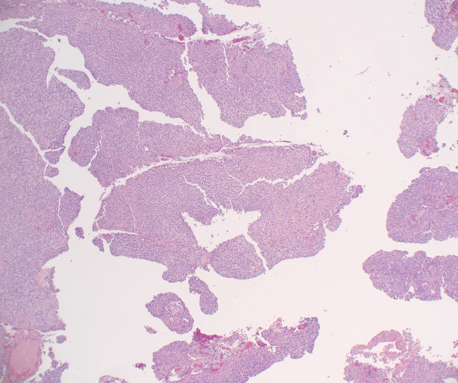
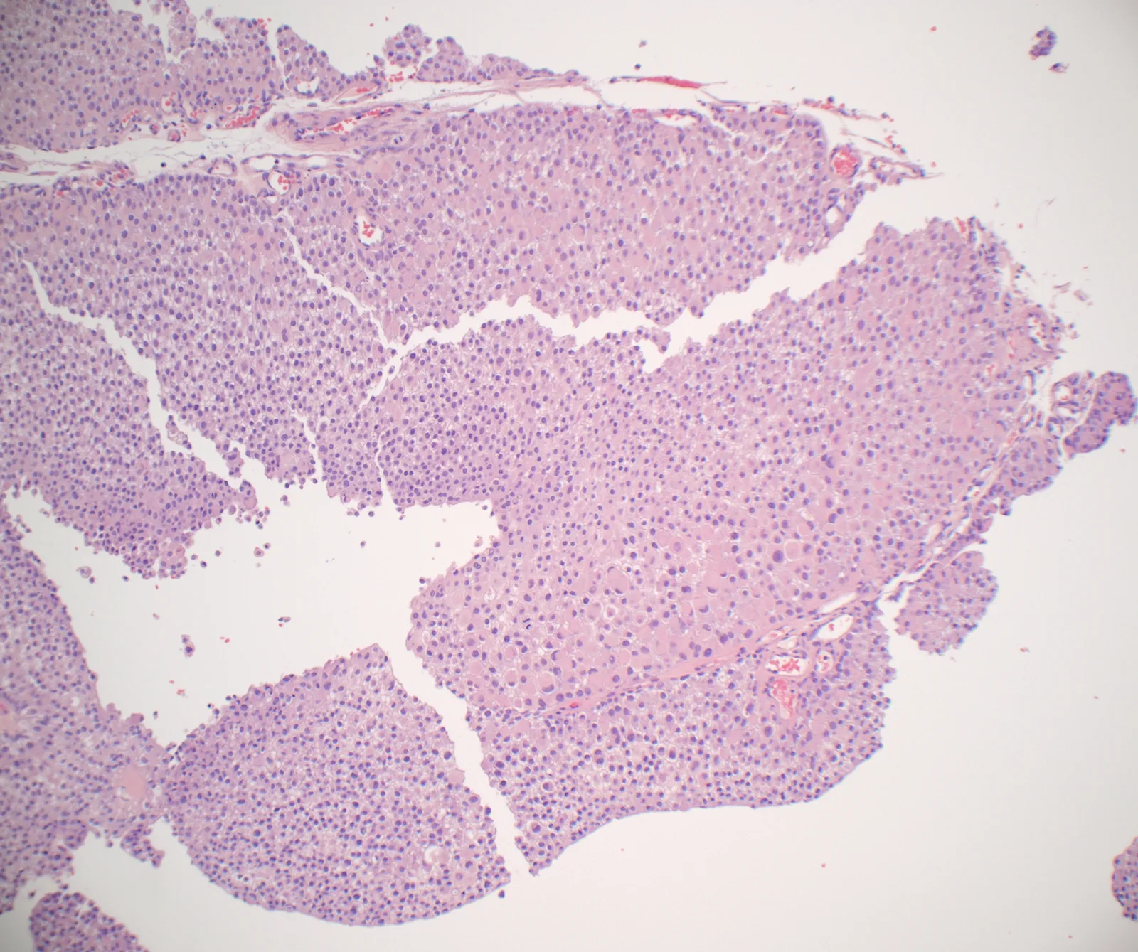
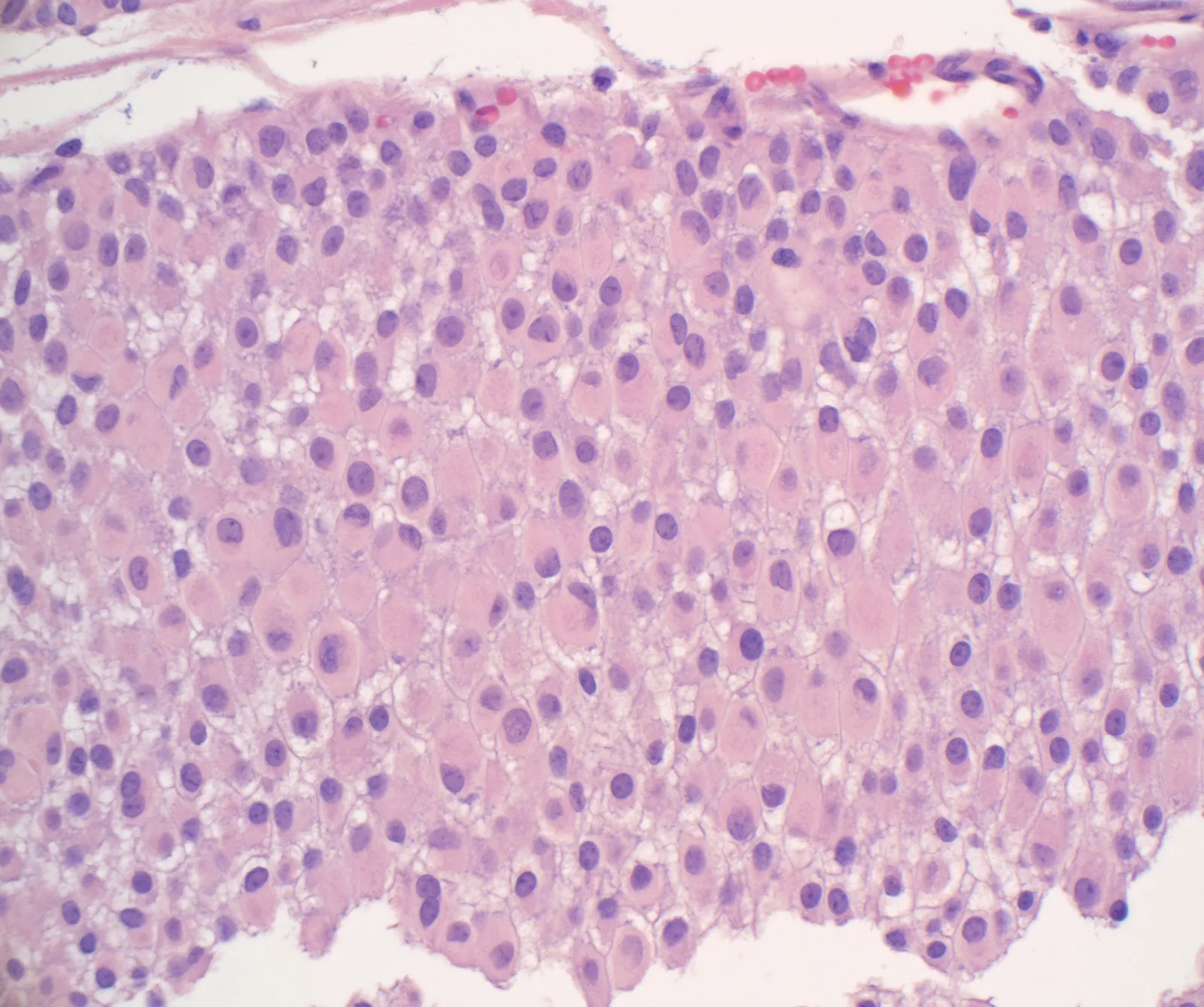
- A pTa papillary urothelial carcinoma at TURBT with "plasmacytoid" foci (eccentric nuclei, eosinophilic globular cytoplasm) and no invasion: low or high grade in a 2-tier system? Grading of such cases is inconsistent in practice.
- Among 500 consecutive pTa specimens, 22 (3.4%) from 17 patients showed this morphology, comprising 5%–95% of the tumor (mean 60.6%); cases with WHO 2004 high-grade cytology, CIS, invasion or prior invasive carcinoma were excluded.
- Of 15 patients followed (mean 18.1 months), 2 progressed (13.3%): one to pT1 at 1.9 months, one with liver metastasis at 30.2 months.
- Authors: uncommon, but a subset may progress; whether it is low, high or intermediate grade needs further study. Subscription article — abstract plus three H&E images from the corresponding author's X post.
Scope: the PubMed abstract, plus the corresponding author's X post and its three images. The article is SAGE subscription content (not OA per Unpaywall); the full text and the article figures were not obtained.
Abstract
We have anecdotally encountered non-invasive (pTa) papillary urothelial carcinomas (PUCs) with eccentric nuclear localization and eosinophilic globular cytoplasm, imparting a "plasmacytoid" appearance, with inconsistent grading in clinical practice. These tumors lack conventional high-grade cytologic features, but show concerning architectural disorder for low grade; in some, associated subsequent invasive carcinoma was identified. Herein, we investigate the frequency of such pTa PUC, their progression rate, and other adverse subtype histology. A retrospective review of 500 consecutive specimens of transurethral resections/biopsies of pTa PUC was performed and evaluated for the presence of "plasmacytoid" morphology. Clinical follow-up was performed. Exclusion criteria included any pTa PUC containing World Health Organization 2004 high-grade cytology, concurrent carcinoma in situ, concurrent invasion, or any prior invasive urothelial carcinoma. Twenty-two of 500 pTa PUC containing plasmacytoid morphology were identified (rate of 3.4%) from 17 patients with a mean age of 67 years. The extent of plasmacytoid morphology ranged between 5% and 95% (mean = 60.6%). Follow-up was available for 15/17 patients (range 3.1-40.1 months; mean = 18.1 months). Subsequent intravesical treatments included: chemotherapy (n = 7), BCG (n = 4), chemotherapy and BCG (n = 2), surveillance (n = 2), immunotherapy/BCG (n = 1), BCG/immunotherapy/chemotherapy (n = 1). Two of 15 patients showed disease progression (13.3%), one progressing to pT1 (at 1.9 months) and one who developed liver metastasis (at 30.2 months). While pTa PUC with "plasmacytoid" morphology is uncommon, a subset may show disease progression, including metastasis. Further studies are needed to establish whether "plasmacytoid" morphology in pTa PUC should be considered low versus high grade or an intermediate grade.
Corresponding author's X post and images
Corresponding author Ankur Sangoi (@slusagar) posted on X on 2026-11-03: "you get a #GUpath TURBT pTa papillary urothelial carcinoma, with these striking "plasmacytoid" looking foci but no invasion — in 2-tier grading: low vs high🤨⁉️ @KukMariya and I recently explored this in a series PMID: 42517751." The post carried three H&E images and a screenshot of the article's first page (the abstract above; not reproduced). No legends were given; the captions below briefly describe what is shown.