- 两例脊柱硬膜内髓外单发强化肿块(64 岁男性颈段、59 岁女性胸段),影像均倾向脑膜瘤;1 例曾诊断为转移性脑膜瘤(区域颈部淋巴结转移、多次复发),1 例曾诊断为脑膜瘤并复发再切。
- 形态两例迥异:例 1 为单一圆形细胞、胞质嗜双色至透亮;例 2 以假血管瘤样结构为主,局灶分叶、合体样胞质。均无脑膜上皮漩涡;Ki-67 20%–30%,核分裂 7–14/10 HPF。
- 免疫组化:vimentin、CD99 弥漫阳性,EMA 斑片阳性,desmin 局灶阳性;PR、SSTR2 基本阴性(不支持脑膜瘤);例 1 另有广谱角蛋白/Cam5.2 斑片强阳性。RNA 测序:EWSR1::CREM(例 1)、EWSR1::ATF1(例 2)。
- 甲基化陷阱:DKFZ v12.8 无 ICMT 类别,例 2 低置信度匹配脑膜瘤;NCI/Bethesda v2.0 分别提示 ICMT 亚型 A、B。因位于颅外,最终诊断「CNS 间叶性肿瘤伴 FET::CREB 融合,NEC」。脊柱 ICMT 约占已报道病例 6%,作者认为 WHO 的「颅内」要求未来或可取消。
收录范围:PMC 开放获取全文(CC BY-NC-ND 4.0)——临床病史与影像、病理所见、讨论、作者贡献,图 1–2 及图注中英对照;该病例 Letter 无摘要;数据可用性声明与参考文献未收录(正文保留文献序号)。
临床病史与影像
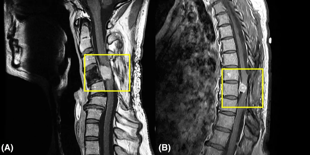
本文报告两例脊柱硬膜内髓外肿块患者。患者 #1 为 64 岁男性,以淋巴结肿大就诊,发现颈椎硬膜内髓外肿块。淋巴结肿大证实为转移性病变,此后多次局部及远处复发,需多次手术。值得注意的是,全身影像检查除脊柱肿块和颈部淋巴结肿大外未见其他病灶。该肿瘤最初倾向诊断为转移性脑膜瘤。患者 #2 为 59 岁女性,以行走困难和感觉异常就诊,发现胸椎硬膜内髓外肿块。肿块切除后当时诊断为脑膜瘤。数年后肿瘤复发并再次切除。两例患者就诊时,脊柱磁共振成像(MRI)均显示单发强化病灶,放射科倾向诊断为脑膜瘤(图 1)。
病理所见
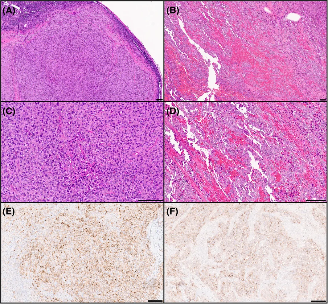
组织病理学检查显示两例患者的形态差异很大,但同一患者各次复发之间的形态学和免疫组化表现相当一致。患者 1 的肿瘤转移至区域颈部淋巴结,由形态单一的细胞构成,核圆形,染色质细颗粒状,核仁小,胞质嗜双色性至透亮(图 2A、C)。患者 2 的肿瘤大部分呈假血管样结构,伴中度异型性,较局灶区域呈分叶状生长、胞质合体样(图 2B、D)。两例均未见脑膜上皮漩涡。Ki-67 增殖指数为 20%–30%,核分裂为每 10 个连续高倍视野 7–14 个。免疫组化:两例均弥漫表达 vimentin 和 CD99,斑片状表达 EMA(图 2F),局灶表达 desmin。患者 1 的肿瘤另外斑片状强阳性表达广谱角蛋白(图 2E)和 Cam5.2。两例肿瘤孕激素受体和 SSTR2 基本均阴性。此外,GFAP、OLIG2、突触素(synaptophysin)、INSM1、抑制素(inhibin)、钙视网膜蛋白(calretinin)和 SF1 均阴性。全转录组 RNA 测序显示患者 #1 的肿瘤存在 EWSR1::CREM 融合,患者 #2 的肿瘤存在 EWSR1::ATF1 融合。
患者 #1 的 DNA 甲基化芯片分类在 DKFZ v12.8 和 NCI/Bethesda v2.0 分类器上均无匹配类别,但 NCI/Bethesda 的部分子分析及 UMAP 嵌入提示颅内间叶性肿瘤 A 亚型。患者 #2 的肿瘤在 DKFZ v12.8 分类器上低置信度匹配脑膜瘤,在 NCI/Bethesda v2.0 分类器上中等置信度匹配颅内间叶性肿瘤 B 亚型。两例最终均诊断为「中枢神经系统间叶性肿瘤,伴 FET::CREB 融合,未归类于他处(NEC)」。采用 NEC 是因为肿瘤位于颅外。
讨论
伴 FET::CREB 融合的颅内间叶性肿瘤是中枢神经系统(CNS)罕见肿瘤,既往曾被称为「颅内血管瘤样纤维组织细胞瘤」或「颅内黏液样间叶性肿瘤」。该肿瘤类型现称为颅内间叶性肿瘤,FET::CREB 融合阳性(ICMT),在 2021 年版 WHO 中枢神经系统肿瘤分类的「间叶性、非脑膜上皮性肿瘤」部分作为暂定实体引入 [1]。其形态学模式多样,常包括血管瘤样、上皮样或黏液样。免疫组化表型常无特异性,已报道 desmin、CD99 和 EMA 呈不同程度阳性。
FET::CREB 融合见于多种软组织肿瘤实体。其发病机制涉及 FET 家族 RNA 结合蛋白(EWSR1 或 FUS)与 cAMP 反应元件结合蛋白(CREB)家族转录因子(如 ATF1、CREB1 和 CREM,三者功能相关)之间的框内基因融合。CREB 信号在神经系统发育和神经可塑性等多种生理过程中发挥重要作用 [2]。这些融合在肿瘤发生中的作用尚不完全清楚。CREB 在包括神经系统发育在内的多种生理过程中起重要作用,更重要的是,其激活与神经可塑性直接相关,而神经可塑性有助于学习和记忆处理 [3]。
目前,2021 年 WHO 分类规定的必要诊断标准之一是 ICMT 位于颅内(幕上或幕下)。然而,也有其他关于颅外 ICMT 的罕见报道。Sloan 等 [4] 对 20 例 FET::CREB 融合肿瘤进行了全面研究,其中 19 例位于脑实质内或脑室内,1 例位于脊髓。该脊髓病例为 15 岁女性,胸段肿瘤,伴 EWRS1::CREM 融合;该患者出现局部复发。同样,Tauziède-Espariat 等报道了 1 例 35 岁女性的颈段脊柱 ICMT,伴 EWSR1::CREM 融合 [5];Kim 等和 Seo 等也报道了伴相同融合的胸段脊柱 ICMT [6, 7]。近期一项 ICMT 大宗病例研究同样指出该实体罕见发生于脊柱,约占已识别病例的 6% [8]。
既往报道的颅外 ICMT 表现为颈段和胸段的轴外强化肿块。多数病例的组织病理学特征以上皮样形态为主,在富于黏液/黏液样的背景中可见星芒状至梭形细胞排列成条索和簇状。既往三例的肿瘤细胞 EMA 和 CD99 阳性,desmin 染色不定。EMA 表达有时会导致误诊为脑膜瘤。DKFZ 甲基化分类器(截至 12.8 版)中没有 ICMT 类别,这类肿瘤常以不同置信度错误匹配到脑膜瘤类别,进一步加剧了与脑膜瘤鉴别的诊断难题。
本文再报告两例颅外「颅内」间叶性肿瘤,凸显了当 ICMT 发生于颅外部位并呈不典型形态时诊断的困难。这些病例有助于认识 ICMT 可发生于整个神经轴,未来版本的 WHO 分类可能无需将「颅内」作为诊断要求。
作者贡献
S. Patel 分析数据并撰写初稿。D Marker 和 T Pearce 分析数据并审阅文章。全体作者批准了文章终稿。
- Two solitary contrast-enhancing intradural extramedullary spinal masses (cervical, 64-year-old man; thoracic, 59-year-old woman) were favored as meningioma on imaging; one was initially called metastatic meningioma (regional cervical nodal metastasis, multiple recurrences), the other diagnosed as meningioma and re-resected on recurrence.
- Morphology differed: case 1 monotonous round cells with amphophilic to clear cytoplasm; case 2 predominantly pseudovascular with focal lobular growth and syncytial cytoplasm. No meningothelial whorls; Ki-67 20%–30%, 7–14 mitoses/10 HPF.
- IHC: diffuse vimentin and CD99, patchy EMA, focal desmin; PR and SSTR2 essentially negative; case 1 also patchy strong pankeratin/Cam5.2. RNA sequencing: EWSR1::CREM (case 1), EWSR1::ATF1 (case 2).
- Methylation pitfall: DKFZ v12.8 lacks an ICMT class (case 2 low-confidence meningioma match); NCI/Bethesda v2.0 pointed to ICMT subtypes A and B. Final diagnosis: CNS mesenchymal tumor with FET::CREB fusion, NEC (extracranial site). Spinal cases are ~6% of reported ICMTs; the authors suggest the intracranial requirement may be unnecessary.
Scope: PMC open-access full text (CC BY-NC-ND 4.0) — clinical history and imaging, findings, discussion and author contributions, with Figures 1–2. This case letter has no abstract; the data availability statement and references are not included (citation numbers are retained in the text).
CLINICAL HISTORY AND IMAGING
Here, we report two patients with intradural extramedullary masses in the spine. Patient #1 is a 64‐year‐old male who presented with lymphadenopathy and was found to have an intradural extramedullary mass in the cervical spine. The lymphadenopathy was confirmed to be metastatic disease, and he experienced multiple episodes of local and distant recurrence, requiring multiple surgeries. Notably, systemic imaging was negative for additional lesions outside of the spinal mass and cervical lymphadenopathy. The tumor was initially favored to be a metastatic meningioma. Patient #2 is a 59‐year‐old female who presented with difficulties walking and paresthesia. She was found to have an intradural extramedullary mass in the thoracic spine. The mass was resected, and a diagnosis of meningioma was issued at the time. The tumor recurred a few years later and was re‐resected. At presentation for both patients, magnetic resonance imaging (MRI) of the spine showed solitary contrast enhancing lesions which were favored to be meningiomas by radiology (Figure 1).
FINDINGS
Histopathologic examination revealed very different morphology between the two patients, but the morphologic and immunohistochemical findings were fairly consistent between recurrences. For patient 1, the neoplasm was metastatic to a regional cervical lymph node and was composed of monotonous cells with round nuclei, finely granular chromatin, and small nucleoli with amphophilic to clear cytoplasm (Figure 2A,C). For patient 2, much of the tumor showed pseudovascular architecture with moderate atypia, with more focal areas of more lobular growth and syncytial cytoplasm (Figure 2B,D). Meningothelial whorls were not seen in either case. The Ki‐67 proliferation indices ranged from 20% to 30%, with mitotic rates between 7 and 14 mitoses in 10 consecutive high‐powered fields. On immunohistochemistry, both cases showed diffuse positivity for vimentin and CD99, patchy positivity for EMA (Figure 2F), and focal positivity for desmin. The tumor from patient 1 additionally showed strong patchy positivity for pancytokeratin (Figure 2E) and Cam5.2. The neoplasms were both essentially negative for progesterone receptor and SSTR2. Additionally, the tumors were negative for GFAP, OLIG2, synaptophysin, INSM1, inhibin, calretinin, and SF1. Whole‐transcript RNA sequencing demonstrated the presence of an EWSR1::CREM fusion in the tumor from patient #1 and an EWSR1::ATF1 fusion in the tumor from patient #2.
DNA methylation array classification for patient #1 showed no matching classes on the DKFZ v12.8 or NCI/Bethesda v2.0 classifiers, but some of the NCI/Bethesda subanalyses and the UMAP embedding suggested intracranial mesenchymal tumor, subtype A. For patient #2, the tumor showed a low‐confidence match for meningioma on the DKFZ v12.8 classifier and a moderate‐confidence match for intracranial mesenchymal tumor, subtype B, on the NCI/Bethesda v2.0 classifier. A final diagnosis of central nervous system mesenchymal tumor with FET::CREB fusion, not elsewhere classified (NEC), was eventually issued in both cases. The NEC designation was applied due to the extracranial location.
DISCUSSION
Intracranial mesenchymal tumors with FET::CREB fusions are rare neoplasms of the central nervous system (CNS) that have been previously described as “intracranial angiomatous fibrous histiocytoma” or “intracranial myxoid mesenchymal tumor.” This tumor type, now termed intracranial mesenchymal tumor, FET::CREB fusion‐positive (ICMT), was introduced as a provisional entity in the “Mesenchymal, Non‐Meningothelial Tumors” section of the 2021 edition of the WHO Classification of Tumors of the Central Nervous System (CNS) [1]. The morphologic patterns are broad and often include angiomatoid, epithelioid, or myxoid. The immunohistochemical profile is often non‐specific, with variable positivity for desmin, CD99, and EMA reported.
FET::CREB fusions are found in a wide variety of soft tissue tumor entities. The pathogenesis involves in‐frame gene fusions of a FET‐family RNA‐binding protein (EWSR1 or FUS) and a transcription factor from the cAMP response element binding protein (CREB) family, such as ATF1, CREB1, and CREM, which are all functionally related. CREB signaling plays an important role in multiple physiologic processes including the development of the nervous system and neuroplasticity [2]. The role these fusions play in oncogenesis is not entirely clear. CREB plays an important role in multiple physiological processes including the development of the nervous system but more importantly, its activation is directly linked to neuroplasticity which aids in learning and memory processing [3].
Currently, one of the essential diagnostic criteria defined by the 2021 WHO classification is that ICMTs are located intracranially (supratentorial or infratentorial). However, there are other rare reports of extracranial ICMTs. Sloan et al. [4] conducted an extensive study of FET::CREB fusion tumors with 20 patients, of whom 19 were intra‐axial or intraventricular, and one was in the spinal cord. The spinal cord presentation was in a 15‐year‐old female with a thoracic tumor with an EWRS1::CREM fusion; this patient experienced a local recurrence. Similarly, Tauziède‐Espariat et al. presented a cervical spinal ICMT with EWSR1::CREM fusion in a 35‐year‐old female [5], and Kim et al. and Seo et al. presented thoracic spinal ICMTs with the same fusion as well [6, 7]. A recent large series of ICMT also notes rare spinal presentations of this entity, accounting for approximately 6% of identified cases [8].
The previously reported extracranial ICMTs showed extra‐axial cervical and thoracic masses with contrast enhancement. Most cases exhibit histopathologic features characterized by predominantly epithelioid morphology, with cords and clusters of stellate to spindle cells in a mucin/myxoid‐rich background. The tumor cells in the prior three cases were positive for EMA and CD99, with variable staining for desmin. The expression of EMA can sometimes lead to a misdiagnosis of meningioma. The diagnostic dilemma with meningioma is further exacerbated by the absence of ICMT in DFKZ methylation classifier (as of version 12.8), where these tumors often show false matches of variable confidence to meningioma classes.
Here, we present two additional cases of extracranial “intracranial” mesenchymal tumors, which highlight the difficulties in diagnosing ICMT when these tumors present in extracranial locations and exhibit atypical morphologic features. The cases presented here contribute to the understanding that ICMT can occur throughout the neuroaxis and that the intracranial requirement for diagnosis may be unnecessary in future editions of the WHO classification.
AUTHOR CONTRIBUTIONS
S. Patel analyzed the data and wrote the draft. D Marker and T Pearce analyzed the data and reviewed the article. All authors approved the final version of the article.