- Histopathology:8 例低分化肺腺癌同时强表达 ALK 与 CD30,形态酷似 ALK 阳性间变性大细胞淋巴瘤(ALCL)——4 例影像初判淋巴瘤,1 例外院误诊并予 BV‐CHOP 后进展。
- 组织学:间变性大细胞弥漫片状,常含马蹄形/肾形核标志性细胞;上皮标志物与 TTF‐1 阳性,淋巴标志与克隆性 TCR 缺如;ALK 融合伴侣属 LUAD 谱系(如 EML4::ALK、PRKAR1A::ALK)。
- 治疗:误诊者换用 alectinib 后获部分缓解;接受 ALK‐TKI 者多数 PR。CD30 在 ALK 阳性 LUAD 中少见(切除 1.6%、活检 3.9%),几乎均伴间变性大细胞形态。
- 实践要点:EMA/CD30/ALK 三联阳性不足以在小活检定诊 ALCL;必须整合肺肿块、上皮/TTF‐1、淋巴标志阴性与 LUAD 型 ALK 融合,以免耽误一线 ALK‐TKI。
收录范围:PMC 开放获取全文(CC BY-NC-ND 4.0)——摘要、引言、材料与方法、结果、讨论、作者贡献,图 1–3、表 1–2 中英对照;利益冲突声明、补充材料、数据可用性声明与参考文献未收录(正文保留文献序号)。
摘要
尽管 ALK 重排是肺腺癌(LUAD)中已确立的改变,CD30 在此背景下的表达仍知之甚少。我们分析一组同时表达 ALK 与 CD30、形态酷似 ALK 阳性间变性大细胞淋巴瘤(ALCL)的低分化 LUAD,聚焦其临床病理与分子特征及潜在诊断与治疗意义。
我们回顾性检索多中心病例库与已发表文献,最终纳入 8 例同时表达 ALK 与 CD30 的低分化 LUAD。系统记录并分析临床、组织形态、免疫组化与分子特征,以及治疗反应与转归。患者中位年龄 47.5 岁(范围 26–67 岁),男女各半,均呈晚期(III–IV 期)。诊断挑战显著:4 例影像学初判为侵袭性淋巴瘤,1 例外院误诊为 ALCL 并已按该病积极治疗后方转诊。组织学上,全部肿瘤由核仁显著、核分裂活跃的显著多形性大细胞弥漫片状构成,形态酷似 ALCL 标志性细胞。多数病例上皮标志物呈弥漫或局灶阳性,TTF‐1 阳性程度不一。Ki‐67 增殖指数 50%–95%。全部病例 ALK 与 CD30 共表达,缺乏淋巴瘤相关免疫表型标志,且无克隆性 T 细胞受体(TCR)基因重排证据。分子测序证实全部 ALK 融合断点均属既往描述的 LUAD 相关变异,所有患者对 ALK 靶向治疗均获显著反应。
双表达 ALK 与 CD30 的低分化 LUAD 少见但诊断极具挑战,可酷似 ALK 阳性 ALCL。认识这一拟态对确保准确诊断并及时启动恰当靶向治疗至关重要。
引言
肺癌是全球最常见恶性肿瘤之一,也是癌症相关死亡的主要原因,死亡率主要集中于晚期患者。全部肺癌中约 85% 归为非小细胞肺癌(NSCLC),其中肺腺癌(LUAD)约占一半。 1 , 2 , 3 , 4 , 5 LUAD 有六种主要组织学生长方式:贴壁、乳头、腺泡、筛状、实性与微乳头。 4 , 6 依据肿瘤内主导方式及其比例,分为高、中、低分化;筛状、实性或微乳头结构合计超过 20% 者定为低分化。 4 该分级预后较差,远处转移风险明显高于高分化或中分化肿瘤。 4 , 7 免疫组化上,LUAD 特征性表达上皮标志物泛角蛋白、EMA、CK7 和/或 CK8/18,以及谱系标志 TTF‐1 与 Napsin A, 8 , 9 , 10 , 11 , 12 但在低分化变异型中这些标志可显著减弱或缺失,增加病理诊断难度。 9 , 13 分子层面,间变性淋巴瘤激酶(ALK)基因重排是 LUAD 关键致癌驱动之一,见于 3%–5% 患者。 14 典型重排来自 2p(p21–p23)臂内倒位,使 EML4 与 ALK 融合形成 EML4::ALK 癌蛋白。较少见的融合伴侣还包括 KIF5B、KLC1、TPM3、STRN、ASXL2、ATP6V1B1、SPDYA 与 PRKAR1A。 15 , 16 选择性 ALK 酪氨酸激酶抑制剂(TKI)已改变 ALK 阳性 LUAD 的治疗格局,现为该分子亚型的标准治疗。 17
CD30 属肿瘤坏死因子受体(TNFR)超家族成员,是多种淋巴造血肿瘤的既定诊断标志,尤以间变性大细胞淋巴瘤(ALCL)、经典型霍奇金淋巴瘤(cHL)与胚胎性癌为代表。 18 , 19 正常造血细胞中,活化 B、T 淋巴细胞强表达 CD30,活化单核细胞与嗜酸性粒细胞仅弱阳性。 19 , 20 CD30 亦见于部分上皮性实体瘤,包括间皮瘤、鼻咽癌与低分化胃癌。 21 , 22 , 23 在 ALCL 中,无论 ALK 状态几乎全部病例表达 CD30;持续 CD30 信号促进肿瘤细胞存活与增殖并利于免疫逃逸。 20 ALK 阳性 ALCL 主要累及儿童与青年,ALK 阴性 ALCL 更多见于中老年,但二者细胞形态难以区分。 20 , 24 尽管存在小细胞、富组织细胞与霍奇金样等形态变异,ALK 阳性 ALCL 几乎恒定含标志性间变性大细胞。 24 t(2;5)(p23;q35) 易位产生 NPM1‐ALK 融合癌蛋白是主要分子事件;较少见的融合伴侣包括 TPM3、ATIC、TFG、CLTC、MSN、TPM4、MYH9、RYH9 与 RNF213。 25 , 26 , 27 , 28 , 29 抗 CD30 抗体–药物偶联物 brentuximab vedotin(BV)已获批用于 ALCL,并显示显著临床疗效。 18 , 30 , 31 CD30 是否在 LUAD 中表达、是否与 ALK 共现,仍不清楚。
本文报告 8 例以间变性细胞为主的低分化 LUAD,在临床表现、组织形态及 CD30/ALK 双阳性免疫反应上酷似 ALK 阳性 ALCL。通过对这些病例临床病理、形态、免疫组化与分子特征的系统分析,我们希望提高对这一诊断挑战性 LUAD 变异型的认识,降低误分类风险,并支持及时、恰当的治疗。
材料与方法
病例识别与文献回顾
自中国广东四家三级医疗中心病理档案中回顾性检索 2014 年 1 月至 2025 年 10 月诊断的 217 例 ALK 阳性 LUAD,包括 62 例手术切除标本与 155 例活检标本。全部病例复核 CD30 免疫组化,记录中至强阳性者。随后识别并纳入 7 例同时表达 ALK 与 CD30 的低分化 LUAD。补充以题名/摘要字段含「CD30」「ALK」「lung」的 PubMed 检索,另获 1 例符合条件的英文病例报告并提取临床病理资料。ALK 与 CD30 共表达但缺乏上皮标志物或 TTF‐1 表达的病例予以排除。最终研究队列共 8 例:7 例来自机构档案,1 例来自已发表文献。诊断材料分别取自肺(n = 4)、回肠(n = 1)、颈淋巴结(n = 1)、腰椎(n = 1)以及胸椎并同时累及肺(n = 1);回肠标本为手术切除,其余均为空心针活检。系统收集并分析临床病理特征、治疗方案与生存结局。所有患者均签署书面知情同意,研究方案经机构伦理委员会批准(批号 SL‐G2023‐039‐01)。
组织形态与免疫组化分析
全部标本经福尔马林固定、石蜡包埋,切 4 μm 厚片并行苏木精–伊红(H&E)染色。纳入须同时满足以下诊断标准:(1)存在原发肺肿块;(2)上皮标志物(包括泛细胞角蛋白 AE1/AE3、CK7 和/或 CK8/18)强阳性或局灶阳性(病例 8 上皮标志 Claudin 4 与 MOC31 低表达);(3)TTF‐1 强阳性或局灶阳性;(4)CD30 中至强阳性且 ALK(克隆 D5F3)强阳性;(5)缺乏与 ALCL 或其他淋巴瘤亚型相关的免疫表型标志;(6)无克隆性 T 细胞受体基因重排(TCR)证据。全部病例由两位病理医师独立复阅;组织学评估涵盖结构生长方式、胞质特征、核形态、核分裂活性、肿瘤坏死与淋巴浆细胞浸润。每次免疫组化均设同步阳/阴性对照,抗体克隆、来源与工作稀释度详见补充表 S1。
靶向二代测序(NGS)
自福尔马林固定石蜡包埋(FFPE)肿瘤标本提取基因组 DNA,采用 OncoScreen Plus 面板(520 个癌症相关基因;1.64 Mb)行靶向 NGS。文库构建、杂交捕获与扩增按厂家方案进行。文库于 Illumina NextSeq 500 平台双端测序,平均测序深度约 1000×。
结果
临床病理特征
全部 8 例患者的临床资料(含文献病例 8 32 )汇总于表 1。队列含男 4 例、女 4 例,中位年龄 47.5 岁(范围 26–67 岁)。首发症状多样,包括胸痛、咳嗽、颈淋巴结肿大、腰痛,以及伴肠转移者的肠梗阻或消化道出血等。胸部 CT 均见肺肿块;8 例中 7 例存在肺癌常见转移区淋巴结肿大(纵隔、肺门或锁骨上)。远处转移常见:骨受累(髂骨、胸骨和/或腰椎)4 例,胃肠道转移(小肠、胃或胰腺)3 例。全部为 III–IV 期。7 例无既往肿瘤史;第 8 例病史不详。4 例影像学初判为淋巴瘤。病例 2 外院误诊为 ALCL,予 4 周期 BV 联合环磷酰胺、多柔比星、长春新碱与泼尼松(BV‐CHOP)后疾病进展;修订诊断为低分化 LUAD 后改用 alectinib,症状显著改善。
| 病例 a | 年龄/性别 | 吸烟 | 临床表现 | 部位 | 治疗 | 反应 | 原发灶大小 | 随访(月) | TNM 分期 |
|---|---|---|---|---|---|---|---|---|---|
| 1 | 67/M | Y | 腹痛/腹胀 | 空肠、髂骨、腹盆腔及纵隔/肺门淋巴结 | Alectinib | SD | 3.8 cm | 5,存活 | IVB |
| 2 | 39/F | N | 咳嗽+胸痛 | 肺、胰腺、胸骨/骶骨、肺门淋巴结 | BV + CHOP, alectinib | PD(BV + CHOP), PR | 5.1 cm | 9,存活 | IVB |
| 3 | 52/F | N | 锁骨上淋巴结肿大+胸痛 | 肺、胸膜、肺门/纵隔/锁骨上淋巴结 | Ensartinib | PR | 1.1 cm | 7,存活 | IVB |
| 4 | 48/M | Y | 咳嗽+黑便 | 肺、胃、肠、肺门淋巴结 | Alectinib | PR | 4.5 cm | 38,存活 | IVB |
| 5 | 26/F | N | 颈淋巴结肿大 | 肺、肺门/锁骨上淋巴结 | Crizotinib | PR | 6.8 cm | 45,死亡 | IIIC |
| 6 | 47/M | N | 腰痛 | 肺、腰椎、肺门淋巴结 | Crizotinib | PR | 6.0 cm | 36,死亡 | IVB |
| 7 | 39/F | N | 干咳 | 肺、纵隔/锁骨上淋巴结 | Lorlatinib+AP | PR | 7.7 cm | 18,存活 | IVA |
| 8 a | 64/M | N | 咳嗽+背痛 | 纵隔/肺门、腰椎 | Alectinib | PR | 10 cm | NA | IV |
AP,阿霉素+顺铂;F,女;LN,淋巴结;M,男;N,否;NA,不可得;NED,无瘤生存;PD,疾病进展;PR,部分缓解;SD,疾病稳定;Y,是。
PMID: 38032068。
组织病理学所见
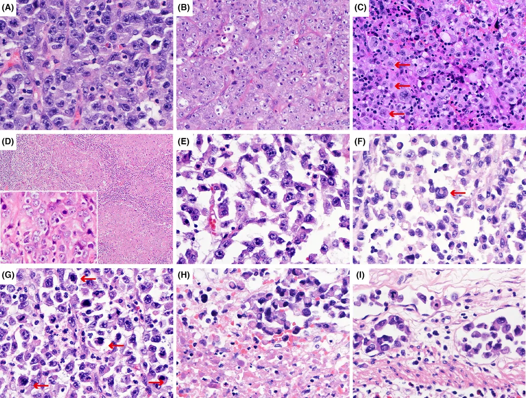
全部病例呈间变性大细胞弥漫增殖;仅病例 8 另见局灶乳头状结构。肿瘤细胞均一显示空泡状核、核仁显著并常见双核(图 1A),病例 3、5、7 空泡状核尤为突出(图 1B)。病例 4 以核大小显著多形(小、中、大核并存)为特征(图 1C)。病例 7 转移淋巴结中肿瘤细胞呈以窦内为主的片状排列(图 1D)。病例 1、6 可见局灶细胞黏附性丧失(图 1E);多例可见偏心马蹄形或肾形核的标志性细胞(图 1F)。7 例可评估病例核分裂 3–12 /mm2(图 1G),其中 4 例(病例 1、2、4、5)见肿瘤坏死(图 1H)。唯一手术切除标本病例 1 尚见广泛脉管侵犯,表现为血管内瘤栓(图 1I)。
免疫表型
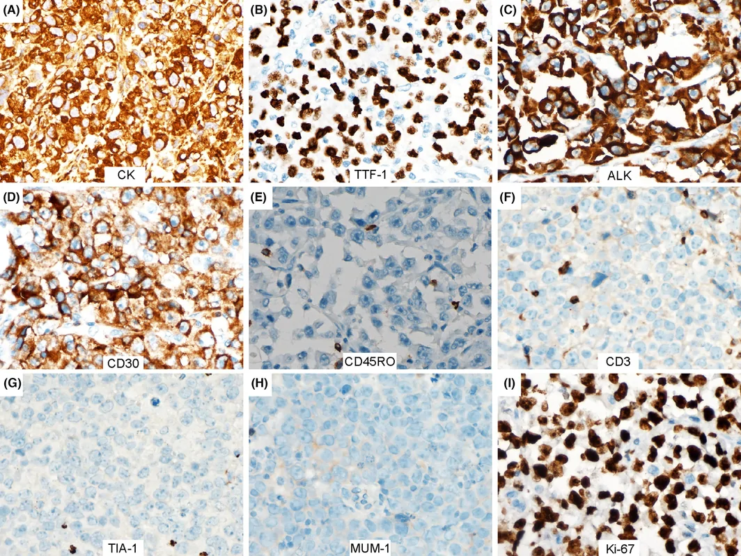
免疫组化结果汇总于表 2。全部病例一种或多种上皮标志物(泛细胞角蛋白 AE1/AE3、CK7 和/或 CK8/18)阳性,呈强或局灶染色(图 2A)。TTF‐1 全部强阳性或局灶阳性(图 2B);Napsin A 见于病例 2、7。全部病例 ALK 与 CD30 强共表达(图 2C,D)。7 例检测 CD45RO 均阴性(图 2E),无一例表达 T 或 B 细胞相关标志(包括 CD2、CD3、CD4、CD5、CD20、CD25、CD43、CD45、MUM‐1、TIA‐1、Perforin 与 granzyme B)(图 2F,G,H;表 2)。7 例可评估病例 Ki‐67 增殖指数 50%–95%(图 2I;病例 8 数据不可得)。3 例评估 PD‐L1 者(病例 1、5、7)均高表达(肿瘤比例评分 >50%)(表 2)。
| 病例 a | CK(%) | CK7(%) | CK8/18 | TTF‐1(%) | Ki67(%) | ALK | CD30(%) | CD2 | CD3 | CD4 | CD5 | CD20 | CD25 | CD43 | CD45RO | MUM1 | TIA‐1 | NapsinA | CD45 | PD‐L1(%) | Grb | Perforin |
|---|---|---|---|---|---|---|---|---|---|---|---|---|---|---|---|---|---|---|---|---|---|---|
| 1 | 10 | NA | NA | ++ | 95 | ++ | ++ | − | − | − | − | − | − | − | − | − | − | − | − | 70 | − | − |
| 2 | 30 | NA | NA | ++ | 50 | ++ | ++ | − | − | − | NA | − | − | − | − | NA | − | 50 | − | NA | NA | NA |
| 3 | 30 | NA | 40 | ++ | 70 | ++ | ++ | NA | − | − | NA | − | − | − | − | − | − | − | − | NA | − | − |
| 4 | 20 | 50 | 60 | 10 | 80 | ++ | ++ | − | − | NA | − | − | NA | − | − | − | − | − | − | NA | − | − |
| 5 | − | 70 | NA | ++ | 85 | ++ | ++ | − | − | − | − | − | − | − | − | − | − | − | − | 65 | − | − |
| 6 | 40 | NA | 50 | 40 | 50 | ++ | ++ | − | − | − | − | − | − | NA | − | − | − | − | − | NA | − | − |
| 7 | ++ | ++ | ++ | ++ | 90 | ++ | ++ | − | − | − | − | − | − | − | − | − | − | ++ | − | ++ | − | − |
| 8 a | − | − | NA | 60 | NA | ++ | ++ | − | − | − | − | − | − | − | NA | − | − | − | − | NA | − | NA |
++,阳性细胞 >80%;− 表示阴性;ALK,ALK(D5F3)。
Grb,granzyme B;NA,不可得。
PMID: 38032068。
分子发现
病例 1、7、8 行靶向 NGS,结果见补充表 S2。病例 1 携带 TP53、BRINP3、NOTCH3 与 ERBB3 体细胞突变(VAF 分别为 42.39%、18.29%、11.64% 与 9.66%),MYC 与 NFKBIA 拷贝数扩增,以及 I 类 PRKAR1A::ALK 融合。病例 7 携带 ARID1B、CREBBP、ATM、AMER1、CSF1R、KMT2C、H3‐3B、NOTCH2、SIPA1、WRN、COL1A1 与 TP53 突变(VAF 3.21%–9.41%),MYC 扩增及 I 类 EML4::ALK 融合。病例 8 携带 NF1 与 TP53 突变及典型 EML4::ALK 融合。 32 病例 1 与 7 均为低肿瘤突变负荷(TMB‐L)、微卫星稳定(MSS),均未检出胚系改变。病例 3、4、6 经荧光原位杂交(FISH)独立证实 ALK 重排。病例 1–7 及既往报告的病例 8 均呈多克隆 TCR 格局。 32
治疗与临床转归
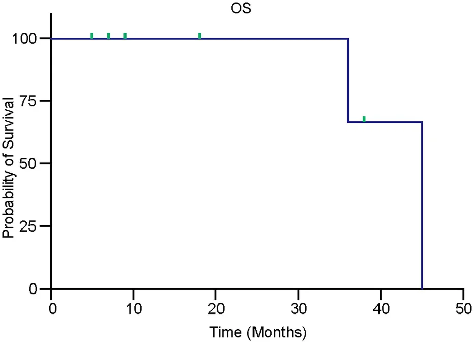
治疗与转归汇总于表 1。全部患者为晚期(III–IV 期),8 例中 7 例未行手术切除。病例 1 因肠梗阻行小肠切除,原发肺病灶留置于原位;随后接受 alectinib 达疾病稳定。病例 2 外院误诊为 ALCL,予 4 周期 BV‐CHOP 后进展;修订为低分化 LUAD 后启用 alectinib,获部分缓解。接受系统治疗的 7 例中 6 例获部分缓解。主要治疗方式为 ALK 靶向:5 例接受 alectinib、ensartinib 或 crizotinib,1 例接受 lorlatinib 联合阿霉素与顺铂(表 1);无一例接受免疫治疗。7 例有随访资料(范围 5–45 个月)。病例 1、2、3、7 随访期内无复发。病例 4 于 25 个月复发,末次随访仍存活。病例 5 于 12 个月进展、45 个月死亡;病例 6 于 11 个月进展、36 个月死亡(图 3)。
CD30 表达在 ALK 阳性 LUAD 中少见
62 例手术切除的 ALK 阳性 LUAD 中 1 例 CD30 阳性(1.6%;病例 1),155 例 ALK 阳性活检中 6 例阳性(3.9%;病例 2–7)。总体而言,ALK 重排 LUAD 中 CD30 表达不常见。全部 7 例 ALK+/CD30+ 病例均为晚期(III–IV 期)并呈间变性大细胞形态。ALK+/CD30+ 与 ALK+/CD30− 肿瘤临床病理比较见补充表 S3。两组在疾病分期(P = 0.034)、组织学亚型(P = 0.010)、大细胞形态(P < 0.001)、Ki‐67 增殖指数(P = 0.002)与 PD‐L1 表达(P < 0.001)上差异有统计学意义。
讨论
尽管 LUAD 中异常 ALK 表达与基因重排已有充分记载,CD30 与 ALK 共表达仍基本未被刻画。据我们所知,本研究是对该现象较早的系统分析之一。通过整合临床、影像、组织形态、免疫组化与分子数据,我们的发现拓展了低分化 LUAD 的形态与免疫表型谱,并界定了一类具有直接治疗相关性的诊断挑战亚群。
CD30/ALK 双阳性 LUAD 是少见但临床后果严重的 ALK 阳性 ALCL 拟态。本队列全部患者以晚期起病,伴广泛淋巴结肿大、远处器官受累及多灶溶骨等结外表现,这些发现合在一起自然首先令人怀疑淋巴瘤。组织学上弥漫片状核仁显著的大间变性细胞及偏心马蹄形或肾形核标志性细胞,免疫表型上 CD30 与 ALK 双阳性,进一步强化了这一印象。 33 尽管存在上述重叠,若干特征始终更支持低分化 LUAD。人口学上,ALK 阳性 ALCL 主要累及儿童与青年,常伴全身 B 症状(发热、盗汗、体重下降);而本队列涵盖青年、中年与老年,且缺乏此类症状。 34 , 35 影像学上,全部病例均有主导性肺肿块伴肺门淋巴结肿大,与极为罕见的原发肺 ALCL 不符。 36 形态学上,全部病例以酷似间变性大细胞淋巴瘤的间变性大细胞为主,凸显辅助检测不可或缺。 35 免疫表型方面,尽管多数 ALK 阳性 ALCL 表达上皮膜抗原(EMA), 37 , 38 且部分病例亦有角蛋白表达报道, 39 , 40 但检出上皮标志物尚不足以据此诊断低分化癌。 40 , 41 本系列中上皮标志物(包括泛角蛋白、CK7 与 CK8/18)弥漫或局灶表达,以及相对 LUAD 特异的 TTF‐1,加上细胞毒性分子与 B/T 细胞相关抗原缺如,支持非淋巴造血起源。分子上,ALK 阳性 ALCL 以 NPM1 为主要 ALK 融合伴侣,而 ALK 阳性 LUAD 以 EML4 及其他 LUAD 相关基因为主 15 , 16 , 25 , 29 ;三例经分子分析的病例(病例 1、7、8)所鉴定融合伴侣均属 LUAD 相关谱系。此外,全部病例 TCR 基因重排分析均无支持淋巴瘤的证据。鉴于 TCR 重排分析可有假阳性与假阴性,且 ALK 阳性 ALCL 常表达 EMA, 37 , 38 在全身淋巴结肿大患者的小活检中,EMA、CD30 与 ALK 同时阳性尚不足以确诊 ALK 阳性 ALCL——尤其在缺乏令人信服的 T 细胞谱系证据时。当背景淋巴细胞混杂表达 T 细胞标志时,诊断挑战更大。因此,准确分类需要综合临床、影像、形态、免疫表型与分子发现。
CD30 是 ALCL 与经典型霍奇金淋巴瘤的既定诊断标志,但其在包括肺癌在内的上皮性恶性肿瘤中的表达仅偶有记载。2014 年 ASCO 年会的一项探索性分析在 17 例 ALK 阴性与 21 例 ALK 阳性 NSCLC 标本中均未检出 CD30 免疫反应。 42 与之相符,我们的数据提示 CD30 表达在 ALK 重排 LUAD 中并不常见。值得注意的是,本队列全部 CD30 阳性肿瘤均呈间变性大细胞形态;无间变性形态的 ALK 阳性 LUAD 中 CD30 阳性率更低。尽管既往系列未描述形态特征, 42 这一观察或可部分解释该系列中未见 CD30 表达。此外,若干肺癌细胞系(H460、H358、A549、H1666 与 H1299)实验上已显示表达 CD30。 43 这一差异可能反映生物学异质性、体外模型作为临床组织替代的固有局限,以及 NSCLC 中 CD30 表达确实罕见。尽管临床数据大体阴性,仍有散在报道记载肺癌及其他上皮性恶性肿瘤的 CD30 表达。在肺癌中,Siba El Hussein 等 描述一例共表达 CD30 与 ALK、形态与免疫表型酷似 ALCL 的低分化 LUAD,情景与本文病例直接类似。 32 肺以外,Grethlein 等 报告一例具间变性细胞学特征的 CD30 阳性低分化胃癌, 23 Wakely 与 Chakhachiro 记载未分化非角化性鼻咽癌的 CD30 表达,其肿瘤细胞呈淋巴结窦内浸润,并具明显核增大与核仁显著的间变性形态。 22 , 44 在 ALK 阳性 ALCL 中,CD30 特征性为细胞膜与高尔基区弥漫强阳性。 30 , 45 本队列病例则呈强膜染色。尽管这一差异在取材充分的切除标本中或有助鉴别,但在小活检中极易被忽略——此时强烈的 CD30 阳性可使低分化癌与 ALK 阳性 ALCL 几乎无法区分,而这一区分具有深远治疗后果。综合来看,CD30 表达并非谱系限定,而可能是上皮与淋巴造血恶性肿瘤中与间变性细胞形态相关的特征。因此,CD30 免疫染色应作为整合形态、免疫表型与分子数据的多参数诊断评估的一环,而非单独分类器。
一项纳入 46 例 ALK 阳性 ALCL 的已发表 NGS 分析中,最常突变基因为 TP53、KMT2 与 CLMN(各 11%),其次为 JUNB、GPS2、NTRK3 与 PKD1(各 7%)以及 MDM4、SETD2、TET1 与 PREX2(各 4%);另有 10 个基因(EP300、PRDM1、NOTCH1、ERBB4、ACSBG2、ARHGEF7、JAK3、SOCS6、DSP 与 FLG)各在单例突变(2%)。 46 , 47 在 ALK 阳性 NSCLC 中,Kron 等 报告 216 例患者中 TP53 共突变频率为 23.8%,其他共突变基因包括 MET(5.4%)、HER2(2.9%)、FGFR2(1.8%)、CTNNB1(1.4%)、PTEN(0.7%)、DDR2(0.7%)与 BRAF(0.6%)。 48 本队列三例经测序的 CD30/ALK 双阳性病例均携带 TP53 突变,其余突变基因与 ALK 阳性 ALCL 突变谱无重叠(补充表 S2)。因此 TP53 成为两类肿瘤共有的唯一复发性突变基因,提示尽管突变景观总体分歧,仍可能存在共同分子脆弱性——尚需在更大、测序更全面的队列中证实。
本队列肿瘤生物学侵袭性强,表现为就诊时分期晚、转移负荷高、Ki‐67 指数显著升高。这种侵袭性叠加与 ALK 阳性 ALCL 显著的形态与免疫表型重叠,使准确分类既困难又至关重要,因为二者治疗根本不同。ALK 阳性 ALCL 主要予联合化疗,可联用或不联用抗 CD30 抗体–药物偶联物 BV,ALK‐TKI 仅保留用于复发或难治疾病。 30 , 31 , 49 相反,ALK 阳性 LUAD 以 ALK‐TKI 作为既定一线标准治疗 17 ;误诊为 ALCL 将使患者失去高效靶向治疗并暴露于不当细胞毒治疗。唯一被误诊为 ALCL 的患者在 brentuximab vedotin 联合 CHOP 上进展,而所有接受 ALK‐TKI 为基础治疗的患者均获良好反应,这一对比直接证明正确诊断的治疗重要性。本队列还引出两点治疗思考。其一,ALK‐TKI 耐药后 BV 是否仍有价值,是值得前瞻评估的临床相关问题。其二,病例 1、5、7 的高 PD‐L1 表达与 ALK 阳性 ALCL 及 ALK 阳性 LUAD 中 PD‐L1 升高的报道一致, 24 , 50 , 51 提示精选患者或可考虑免疫检查点抑制,但其在此特定情境中的疗效尚待确立。综上,这些发现凸显一类诊断挑战但治疗上可行动的实体。尽管形态与免疫表型酷似 ALK 阳性 ALCL 且临床病理侵袭,双表达 CD30 与 ALK 的低分化 LUAD 在正确、及时诊断的前提下对 ALK‐TKI 治疗反应良好。
总之,CD30/ALK 双阳性低分化 LUAD 是一种少见但明确的诊断实体,其与 ALK 阳性 ALCL 的形态与免疫表型相似构成胸部病理的重要陷阱。二者治疗根本不同、预后含义亦异,准确识别在临床上不可或缺。正确分类有赖于将临床与影像资料同全面免疫分型(涵盖上皮与淋巴标志)以及 ALK 融合伴侣的分子鉴定相结合。未来研究应阐明 LUAD 中 CD30 表达的生物学基础、其与 TP53 突变等共现改变的相互作用,以及同时靶向 ALK 与 CD30 是否带来治疗获益。
作者贡献
JN 构思并设计研究并总体督导。CWJ、JJT 与 YQ 分析数据并起草文稿。HCY 与 FYF 收集并整理临床病例资料。YQ 与 CYS 完成免疫组化染色与 FISH。全体作者对文稿重要知识内容作了批判性审阅,并批准最终提交版本。
- Histopathology: eight poorly differentiated LUADs with strong dual ALK/CD30 expression closely mimicked ALK-positive ALCL — four were first read as lymphoma on imaging and one was treated as ALCL with BV-CHOP before revision.
- Histology: diffuse anaplastic large cells with hallmark horseshoe/reniform nuclei; epithelial markers and TTF-1 positive, lymphoma markers and clonal TCR absent; ALK fusion partners were LUAD-type (e.g. EML4::ALK, PRKAR1A::ALK).
- Therapy: the misdiagnosed patient responded after switching to alectinib; most patients on ALK-TKIs achieved PR. CD30 is uncommon in ALK-positive LUAD (1.6% resections, 3.9% biopsies) and almost always accompanies anaplastic morphology.
- Practice point: EMA/CD30/ALK co-positivity on a small biopsy is not enough for ALCL — integrate the lung mass, epithelial/TTF-1 profile, negative lymphoid markers and LUAD-type ALK fusion so first-line ALK-TKI is not delayed.
Scope: PMC open-access full text (CC BY-NC-ND 4.0) — abstract, introduction, materials and methods, results, discussion, author contributions, with Figures 1–3 and Tables 1–2. Conflict-of-interest statement, supporting information, data availability statement and references are not included (citation numbers are retained).
Abstract
Although ALK rearrangement is a well‐established alteration in lung adenocarcinoma (LUAD), CD30 expression in this setting remains poorly characterized. We analysed a series of poorly differentiated LUADs with concurrent ALK and CD30 expression that closely resemble ALK‐positive anaplastic large‐cell lymphoma (ALCL), focusing on their clinicopathological and molecular features as well as potential diagnostic and therapeutic implications.
We retrospectively searched multi‐institutional case databases and the published literature, ultimately identifying eight cases of poorly differentiated LUAD with concurrent ALK and CD30 expression. Clinical, histomorphological, immunohistochemical and molecular features, along with treatment responses and outcomes, were systematically recorded and analysed. Patients had a median age of 47.5 years (range, 26–67 years), with equal sex distribution and all presented with advanced‐stage disease (stage III–IV). The diagnostic challenge posed by these tumours was substantial: four cases were initially radiologically suspected to represent aggressive lymphoma, and one patient had been misdiagnosed and actively treated as ALCL at an outside institution before referral. Histologically, all tumours consisted of diffuse sheets of markedly pleomorphic large cells with prominent nucleoli and brisk mitotic activity, a morphology strikingly reminiscent of ALCL hallmark cells. Most cases showed diffuse or focal expression of epithelial markers, with variable TTF‐1 positivity. The Ki‐67 proliferation index ranged from 50% to 95%. All cases co‐expressed ALK and CD30, lacked lymphoma‐associated immunophenotypic markers and showed no evidence of clonal T‐cell receptor gene rearrangement (TCR). Molecular sequencing confirmed that all ALK fusion breakpoints corresponded to previously described LUAD‐associated variants, and all patients achieved marked responses to ALK‐directed therapy.
Poorly differentiated LUAD with dual ALK and CD30 expression is a rare but diagnostically challenging entity that can closely simulate ALK‐positive ALCL. Awareness of this mimic is critical to ensure accurate diagnosis and timely initiation of appropriate targeted therapy.
Introduction
Lung cancer is one of the two most common malignancies worldwide and a leading cause of cancer‐related death, with mortality concentrated predominantly among patients with advanced‐stage disease. Of all lung cancer cases, approximately 85% are classified as non‐small cell lung cancer (NSCLC) and lung adenocarcinoma (LUAD) accounts for roughly half of these. 1 , 2 , 3 , 4 , 5 LUAD exhibits six principal histological growth patterns: lepidic, papillary, acinar, cribriform, solid and micropapillary. 4 , 6 Tumours are graded as well, moderately or poorly differentiated based on the dominant pattern and its proportional extent within the neoplasm; those in which cribriform, solid or micropapillary architecture collectively exceeds 20% are designated poorly differentiated. 4 This grade carries a worse prognosis and a markedly higher risk of distant metastasis relative to well‐ or moderately differentiated tumours. 4 , 7 Immunohistochemically, LUAD characteristically expresses the epithelial markers pan‐keratin, EMA, CK7 and/or CK8/18, together with the lineage‐specific markers TTF‐1 and Napsin A, 8 , 9 , 10 , 11 , 12 although in poorly differentiated variants these markers may be substantially diminished or absent, a finding that can complicate pathological diagnosis. 9 , 13 At the molecular level, anaplastic lymphoma kinase (ALK) gene rearrangements represent one of the key oncogenic driver alterations in LUAD, occurring in 3%–5% of patients. 14 The prototypical rearrangement arises from an intrachromosomal inversion on chromosome 2p (bands p21–p23), fusing EML4 with ALK to generate the EML4::ALK oncoprotein. Additionally, less common fusion partners include KIF5B, KLC1, TPM3, STRN, ASXL2, ATP6V1B1, SPDYA and PRKAR1A. 15 , 16 Selective ALK tyrosine kinase inhibitors (TKIs) have since transformed the management of ALK‐positive LUAD and are now the established standard of care for this molecular subset. 17
CD30, a member of the tumour necrosis factor receptor (TNFR) superfamily, is an established diagnostic marker for several haematolymphoid neoplasms, most notably anaplastic large‐cell lymphoma (ALCL), classical Hodgkin lymphoma (cHL) and embryonal carcinoma 18 , 19 Among normal haematopoietic cells, activated B and T lymphocytes express CD30 strongly, while activated monocytes and eosinophils show only weak positivity. 19 , 20 CD30 expression has additionally been reported in select epithelial solid tumours, including mesothelioma, nasopharyngeal carcinoma and poorly differentiated gastric carcinoma. 21 , 22 , 23 In ALCL, CD30 is expressed in virtually all cases regardless of ALK status, and sustained CD30 signalling promotes tumour cell survival and proliferation while enabling immune evasion. 20 ALK‐positive ALCL predominantly affects children and young adults, whereas ALK‐negative ALCL is more prevalent in middle‐aged and older patients, yet the two subtypes are cytomorphologically indistinguishable. 20 , 24 Despite encompassing morphological variants such as small cell, histiocyte‐rich and Hodgkin‐like patterns, ALK‐positive ALCL invariably contains hallmark anaplastic large cells. 24 The t(2;5)(p23;q35) translocation, generating the NPM1‐ALK fusion oncoprotein, is the predominant molecular event; alternative, less common fusion partners include TPM3, ATIC, TFG, CLTC, MSN, TPM4, MYH9, RYH9 and RNF213. 25 , 26 , 27 , 28 , 29 The anti‐CD30 antibody–drug conjugate brentuximab vedotin (BV) is approved for ALCL and has shown considerable clinical efficacy. 18 , 30 , 31 Whether CD30 is expressed in LUAD and whether it co‐occurs with ALK remains poorly understood.
Herein, we report eight cases of poorly differentiated LUAD composed predominantly of anaplastic cells, which closely mimic ALK‐positive ALCL in clinical presentation, histomorphology and dual CD30/ALK immunoreactivity. Through systematic analysis of the clinicopathological, morphological, immunohistochemical and molecular features of these cases, we seek to raise awareness of this diagnostically challenging LUAD variant, reduce the risk of misclassification and support timely, appropriate treatment.
Materials and methods
Case Identification and Literature Review
A total of 217 ALK‐positive LUADs diagnosed between January 2014 and October 2025 were retrospectively retrieved from the pathology archives of four tertiary medical centres in Guangdong, China, including 62 resection specimens and 155 biopsy specimens. CD30 immunohistochemistry was reviewed in all cases, and tumours showing moderate‐to‐strong CD30 expression were recorded. Seven poorly differentiated LUADs with concurrent ALK and CD30 expression were subsequently identified and included in the study. A complementary PubMed search using the terms ‘CD30’, ‘ALK’ and ‘lung’ in the title and abstract fields identified one additional eligible English‐language case report, and clinicopathological data were subsequently extracted. Cases with ALK and CD30 co‐expression but lacking expression of either epithelial markers or TTF‐1 were excluded from the study. The final study cohort comprised eight cases in total: seven from institutional archives and one from the published literature. Diagnostic material was obtained from the lung (n = 4), ileum (n = 1), cervical lymph node (n = 1), lumbar vertebra (n = 1) and thoracic vertebra with concurrent pulmonary involvement (n = 1); the ileal specimen was surgically resected, whereas all others were obtained by core needle biopsy. Clinicopathological characteristics, treatment regimens and survival outcomes were systematically collected and analysed. Written informed consent was obtained from all patients, and the study protocol was approved by the institutional ethics committee (approval no. SL‐G2023‐039‐01).
Histomorphological and Immunohistochemical Analysis
All specimens were formalin‐fixed, paraffin‐embedded, sectioned at 4 μm and stained with haematoxylin and eosin (H&E). Cases were included if all of the following diagnostic criteria were met: (1) presence of a primary pulmonary mass; (2) strong or focal positivity for epithelial markers, including pan‐cytokeratin (AE1/AE3), CK7 and/or CK8/18 (Case 8 showed low expression of the epithelial markers Claudin 4 and MOC31); (3) strong or focal TTF‐1 expression; (4) moderate‐to‐strong CD30 expression and strong ALK expression (clone D5F3); (5) absence of immunophenotypic markers associated with ALCL or other lymphoma subtypes; (6) no evidence of clonal T‐cell receptor gene rearrangement (TCR). All cases were independently reviewed by two pathologists; histological assessment encompassed architectural growth patterns, cytoplasmic features, nuclear morphology, mitotic activity, tumour necrosis and lymphoplasmacytic infiltration. Each immunohistochemical run was performed with concurrently processed positive and negative controls, and antibody clones, sources and working dilutions are detailed in Table S1.
Targeted Next‐Generation Sequencing (NGS)
Genomic DNA isolated from formalin‐fixed, paraffin‐embedded (FFPE) tumour specimens was processed for targeted NGS via the OncoScreen Plus panel (520 cancer‐associated genes; 1.64 Mb). Library construction, hybridization capture and amplification were carried out in accordance with the manufacturer's protocols. The resulting libraries were sequenced on an Illumina NextSeq 500 platform in paired‐end mode, at a mean sequencing depth of approximately 1000×.
Results
Clinicopathological Characteristics
Clinical data from all eight patients (including Case 8 from the literature 32 ) are summarized in Table 1. The cohort comprised four males and four females with a median age of 47.5 years (range, 26–67 years). Presenting symptoms varied widely, including chest pain, cough, cervical lymphadenopathy, low back pain, and, in patients with bowel metastasis, such as intestinal obstruction or gastrointestinal bleeding. Chest CT revealed a pulmonary mass in all cases, and lymphadenopathy involving regions commonly affected by lung cancer metastasis (mediastinal, hilar or supraclavicular nodal stations) was identified in seven of eight patients. Distant metastases were frequent: osseous involvement (iliac bone, sternum and/or lumbar vertebrae) was documented in four patients and gastrointestinal metastases affecting the small bowel, stomach or pancreas in three. All patients presented with stage III–IV disease. Seven had no prior oncological history; history was unavailable for the eighth. Lymphoma was the initial imaging impression in four cases. Case 2 had been misdiagnosed with ALCL at an outside institution and treated with four cycles of BV plus cyclophosphamide, doxorubicin, vincristine and prednisone (BV‐CHOP), after which the disease progressed. Following diagnostic revision to poorly differentiated LUAD, the patient was switched to alectinib and achieved marked symptomatic improvement.
| Case a | Age/gender | Smoke | Presentation | Location | Treatment | Response | Primary lesion size | Follow‐up (months) | TNM stage |
|---|---|---|---|---|---|---|---|---|---|
| 1 | 67/M | Y | Abdominal pain/distension | Jejunum, iliac bone abdominopelvic & mediastinal/hilar LN | Alectinib | SD | 3.8 cm | 5, Alive | IVB |
| 2 | 39/F | N | Cough+ chest pain | Lung, pancreas, sternum/sacrum, hilar LN | BV + CHOP, alectinib | PD(BV + CHOP), PR | 5.1 cm | 9, Alive | IVB |
| 3 | 52/F | N | Supraclavicular LN enlargement +chest pain | Lung, pleura, hilar /mediastinal/supraclavicular LN | Ensartinib | PR | 1.1 cm | 7, Alive | IVB |
| 4 | 48/M | Y | Cough+ melena | Lung, stomach, intestine, hilar LN | Alectinib | PR | 4.5 cm | 38, Alive | IVB |
| 5 | 26/F | N | Neck LN enlargement | Lung, hilar/supraclavicular LN | Crizotinib | PR | 6.8 cm | 45, Died | IIIC |
| 6 | 47/M | N | Low back pain | Lung, Lumbar vertebrae, hilar LN | Crizotinib | PR | 6.0 cm | 36, Died | IVB |
| 7 | 39/F | N | Dry cough | Lung, mediastinal/supraclavicular LN | Lorlatinib+AP | PR | 7.7 cm | 18, Alive | IVA |
| 8 a | 64/M | N | Cough+ back pain | Mediastinal/hilar, lumbar spinal | Alectinib | PR | 10 cm | NA | IV |
AP, Adriamycin+Cisplatin; F, female; LN, lymph node; M, male; N, no; NA, not available; NED, no evidence of disease; PD, progressive disease; PR, partial response; SD, stable disease; Y, yes.
PMID: 38032068.
Histopathological Findings
All cases showed diffuse proliferation of anaplastic large cells; Case 8 alone also exhibited focal papillary architecture. The tumour cells uniformly displayed vesicular nuclei, conspicuous nucleoli and frequent binucleation (Figure 1A), with vesicular nuclei being especially prominent in Cases 3, 5 and 7 (Figure 1B). Case 4 was distinguished by marked nuclear pleomorphism spanning small, intermediate and large forms (Figure 1C). In the metastatic lymph nodes of Case 7, tumour cells adopted a predominantly sinus‐based, sheet‐like arrangement (Figure 1D). Focal cellular discohesion was noted in Cases 1 and 6 (Figure 1E), and characteristic hallmark cells with eccentric horseshoe‐shaped or reniform nuclei were identified in multiple cases (Figure 1F). Mitotic figures ranged from 3 to 12 per mm2 across all seven evaluable cases (Figure 1G), and tumour necrosis was present in four of these seven (Cases 1, 2, 4 and 5) (Figure 1H). In Case 1, the sole surgical resection specimen, extensive lymphovascular invasion manifesting as intravascular tumour emboli was also observed (Figure 1I).
Immunophenotypic Profiles
The immunohistochemical findings are summarized in Table 2. All cases were positive for one or more epithelial markers (pan‐cytokeratin (AE1/AE3), CK7 and/or CK8/18), showing strong or focal staining (Figure 2A). TTF‐1 was strongly positive or focally positive in all cases (Figure 2B); Napsin A was expressed in Cases 2 and 7. ALK and CD30 were strongly co‐expressed in all cases (Figure 2C,D). All seven cases tested for CD45RO were negative (Figure 2E), and no case showed expression of T‐ or B‐cell–associated markers, including CD2, CD3, CD4, CD5, CD20, CD25, CD43, CD45, MUM‐1, TIA‐1, Perforin and granzyme B (Figure 2F,G,H; Table 2). The Ki‐67 proliferation index ranged from 50% to 95% in all seven evaluable cases (Figure 2I; data unavailable for Case 8). PD‐L1 expression was high (tumour proportion score >50%) in all three cases in which it was assessed (Cases 1, 5 and 7) (Table 2).
| Case a | CK(%) | CK7 (%) | CK8/18 | TTF‐1 (%) | Ki67 (%) | ALK | CD30 (%) | CD2 | CD3 | CD4 | CD5 | CD20 | CD25 | CD43 | CD45RO | MUM1 | TIA‐1 | NapsinA | CD45 | PD‐L1 (%) | Grb | Perforin |
|---|---|---|---|---|---|---|---|---|---|---|---|---|---|---|---|---|---|---|---|---|---|---|
| 1 | 10 | NA | NA | ++ | 95 | ++ | ++ | − | − | − | − | − | − | − | − | − | − | − | − | 70 | − | − |
| 2 | 30 | NA | NA | ++ | 50 | ++ | ++ | − | − | − | NA | − | − | − | − | NA | − | 50 | − | NA | NA | NA |
| 3 | 30 | NA | 40 | ++ | 70 | ++ | ++ | NA | − | − | NA | − | − | − | − | − | − | − | − | NA | − | − |
| 4 | 20 | 50 | 60 | 10 | 80 | ++ | ++ | − | − | NA | − | − | NA | − | − | − | − | − | − | NA | − | − |
| 5 | − | 70 | NA | ++ | 85 | ++ | ++ | − | − | − | − | − | − | − | − | − | − | − | − | 65 | − | − |
| 6 | 40 | NA | 50 | 40 | 50 | ++ | ++ | − | − | − | − | − | − | NA | − | − | − | − | − | NA | − | − |
| 7 | ++ | ++ | ++ | ++ | 90 | ++ | ++ | − | − | − | − | − | − | − | − | − | − | ++ | − | ++ | − | − |
| 8 a | − | − | NA | 60 | NA | ++ | ++ | − | − | − | − | − | − | − | NA | − | − | − | − | NA | − | NA |
++, positive cells >80%; − indicates negative; ALK, ALK (D5F3).
Grb, granzyme B; NA, not available.
PMID: 38032068.
Molecular Findings
Targeted NGS was performed in Cases 1, 7 and 8; results are presented in Table S2. Case 1 carried somatic mutations in TP53, BRINP3, NOTCH3 and ERBB3 (VAFs: 42.39%, 18.29%, 11.64% and 9.66%, respectively), MYC and NFKBIA copy number amplification and a Class I PRKAR1A::ALK fusion. Case 7 carried mutations in ARID1B, CREBBP, ATM, AMER1, CSF1R, KMT2C, H3‐3B, NOTCH2, SIPA1, WRN, COL1A1 and TP53 (VAFs: 3.21%–9.41%), MYC amplification and a Class I EML4::ALK fusion. Case 8 carried mutations in NF1 and TP53 alongside a canonical EML4::ALK fusion. 32 Both Cases 1 and 7 exhibited low tumour mutation burden (TMB‐L) and microsatellite stability (MSS), with no germline alterations detected in either. ALK rearrangement was independently confirmed by fluorescence in situ hybridization (FISH) in Cases 3, 4 and 6. Cases 1–7 and the previously reported Case 8 all demonstrated a polyclonal TCR pattern. 32
Treatment and Clinical Outcomes
Treatment and outcome data are summarized in Table 1. All patients presented with advanced‐stage disease (stage III–IV), and seven of eight did not undergo surgical resection. Case 1 required small bowel resection for intestinal obstruction, though the primary lung lesion was left in situ; this patient subsequently received alectinib and achieved stable disease. Case 2 had been misdiagnosed as ALCL at an outside institution and treated with four cycles of BV‐CHOP, after which the disease progressed. Following diagnostic revision to poorly differentiated LUAD, alectinib was initiated, and the patient achieved a partial response. Among the seven patients who received systemic therapy, six achieved a partial response. The predominant treatment modality was ALK‐directed therapy: five patients received alectinib, ensartinib or crizotinib, and one received lorlatinib combined with adriamycin and cisplatin (Table 1); no patient received immunotherapy. Follow‐up data were available for seven patients (range, 5–45 months). Four patients (Cases 1, 2, 3 and 7) remained recurrence‐free throughout the follow‐up period. Case 4 developed recurrence at 25 months and was alive at the last follow‐up. Case 5 progressed at 12 months and died at 45 months; Case 6 progressed at 11 months and died at 36 months (Figure 3).
CD30 Expression Is a Rare Event in ALK‐Positive LUAD
CD30 positive expression was identified in 1 of 62 resected ALK‐positive LUADs (1.6%; Case 1) and in 6 of 155 ALK‐positive biopsy specimens (3.9%; Cases 2–7). Overall, CD30 expression was uncommon in ALK‐rearranged LUAD. All seven ALK+/CD30+ cases presented with advanced‐stage disease (stage III–IV) and exhibited anaplastic large‐cell morphology. Comparisons of clinicopathological characteristics between ALK+/CD30+ and ALK+/CD30− tumours are summarized in Table S3. Significant differences were observed between the two groups in disease stage (P = 0.034), histological subtype (P = 0.010), large‐cell morphology (P < 0.001), Ki‐67 proliferation index (P = 0.002) and PD‐L1 expression (P < 0.001).
Discussion
Although aberrant ALK expression and gene rearrangements are well documented in LUAD, concurrent CD30 and ALK co‐expression remains largely uncharacterized. To our knowledge, this study is among the first systematic analyses of this phenomenon. Through integration of clinical, radiological, histomorphological, immunohistochemical and molecular data, our findings expand the morphological and immunophenotypic spectrum of poorly differentiated LUAD and define a diagnostically challenging subset with direct therapeutic relevance.
CD30/ALK double‐positive LUAD is a rare but clinically consequential mimic of ALK‐positive ALCL. In our cohort, all patients presented with advanced‐stage disease accompanied by extensive lymphadenopathy, distant organ involvement and extranodal manifestations such as multifocal osteolytic lesions, findings that collectively and understandably raised an initial suspicion of lymphoma. This impression was further reinforced histologically by diffuse sheets of large anaplastic cells with conspicuous nucleoli and hallmark cells bearing eccentric horseshoe‐shaped or reniform nuclei and immunophenotypically by dual CD30 and ALK expression. 33 Despite this overlap, several features consistently favour poorly differentiated LUAD. Demographically, ALK‐positive ALCL predominantly affects children and young adults and is often associated with systemic B symptoms (fever, night sweats and weight loss). In contrast, our cohort included young, middle‐aged and elderly individuals and lacked such symptoms. 34 , 35 Radiologically, a dominant pulmonary mass with hilar lymphadenopathy, present in all cases, is inconsistent with primary pulmonary ALCL, which is exceedingly rare in any form. 36 Morphologically, all cases were predominantly composed of anaplastic large cells resembling those of anaplastic large‐cell lymphoma, highlighting the indispensable role of ancillary testing. 35 Immunophenotypically, although most ALK‐positive ALCLs express epithelial membrane antigen (EMA), 37 , 38 and keratin expression has also been reported in a subset of cases, 39 , 40 the detection of epithelial markers should not be regarded as sufficient evidence for a diagnosis of poorly differentiated carcinoma. 40 , 41 In our cases, diffuse or focal expression of epithelial markers (including pan‐keratin, CK7 and CK8/18) and the relatively LUAD‐specific marker TTF‐1, together with the absence of cytotoxic molecules and B‐ or T‐cell–associated antigens, supported a non‐haematolymphoid origin. Molecularly, whereas NPM1 is the predominant ALK fusion partner in ALK‐positive ALCL, EML4 and other LUAD‐associated genes predominate in ALK‐positive LUAD 15 , 16 , 25 , 29 ; in all three molecularly analysed cases (Cases 1, 7 and 8), the identified fusion partners fell exclusively within the LUAD‐associated spectrum. Moreover, TCR gene rearrangement analysis in all cases provided no evidence supporting a diagnosis of lymphoma. Given the potential for both false‐positive and false‐negative results in TCR gene rearrangement analysis, together with the frequent expression of epithelial membrane antigen (EMA) in ALK‐positive ALCL, 37 , 38 concurrent positivity for EMA, CD30 and ALK in small biopsy specimens from patients presenting with generalized lymphadenopathy should not be considered sufficient for a definitive diagnosis of ALK‐positive ALCL, particularly in the absence of convincing evidence of T‐cell lineage. The diagnostic challenge becomes even greater when admixed background lymphocytes express T‐cell markers. Therefore, accurate classification requires comprehensive integration of clinical, radiological, morphological, immunophenotypic and molecular findings.
CD30 is an established diagnostic marker for ALCL and classical Hodgkin lymphoma, yet its expression in epithelial malignancies, including lung cancer, has been documented only rarely. An exploratory analysis presented at the 2014 ASCO Annual Meeting identified no CD30 immunoreactivity in 17 ALK‐negative or 21 ALK‐positive NSCLC specimens. 42 Consistent with these findings, our data indicate that CD30 expression is uncommon in ALK‐rearranged LUAD. Notably, all CD30‐positive tumours in our cohort exhibited anaplastic large‐cell morphology. The rate of CD30 expression was even lower among ALK‐positive LUADs without anaplastic morphology. Although the morphological features of the previously reported cases were not described, 42 this observation may partly explain the absence of CD30 expression in that series. Furthermore, several lung cancer cell lines (H460, H358, A549, H1666 and H1299) have been experimentally shown to express CD30. 43 This discrepancy likely reflects a combination of biological heterogeneity, the inherent limitations of in vitro models as surrogates for clinical tissue and the genuine rarity of CD30 expression in NSCLC. Despite these largely negative clinical data, sporadic reports have documented CD30 expression in lung cancer and other epithelial malignancies. In the setting of lung cancer, Siba El Hussein et al. described a poorly differentiated LUAD co‐expressing CD30 and ALK that closely simulated ALCL both morphologically and immunophenotypically, a scenario directly analogous to the cases presented here. 32 Beyond the lung, Grethlein et al. reported a CD30‐positive poorly differentiated gastric carcinoma with anaplastic cytological features, 23 and Wakely and Chakhachiro documented CD30 expression in undifferentiated non‐keratinizing nasopharyngeal carcinoma, where tumour cells exhibited sinusoidal lymph node infiltration and overtly anaplastic morphology with marked nuclear enlargement and prominent nucleoli. 22 , 44 In ALK‐positive ALCL, CD30 is characteristically diffuse and strong, involving the cell membrane and Golgi regions. 30 , 45 The cases in our cohort, by contrast, showed strong membranous staining. Although this difference may aid distinction in resection specimens with adequate sampling, it is easily overlooked in small biopsies, where intense CD30 positivity can render a poorly differentiated carcinoma virtually indistinguishable from ALK‐positive ALCL, a distinction with profound therapeutic consequences. Taken together, these findings suggest that CD30 expression is not lineage‐restricted but may instead be a feature associated with anaplastic cytomorphology across both epithelial and haematolymphoid malignancies. Accordingly, CD30 immunostaining should be interpreted as one component of a multiparametric diagnostic evaluation that incorporates morphological, immunophenotypic and molecular data, rather than as a standalone classifier.
In a published NGS analysis of 46 ALK‐positive ALCL cases, the most frequently mutated genes were TP53, KMT2 and CLMN (each 11%), followed by JUNB, GPS2, NTRK3 and PKD1 (each 7%) and MDM4, SETD2, TET1 and PREX2 (each 4%); ten additional genes (EP300, PRDM1, NOTCH1, ERBB4, ACSBG2, ARHGEF7, JAK3, SOCS6, DSP and FLG) were each mutated in a single case (2%). 46 , 47 In ALK‐positive NSCLC, Kron et al. reported a TP53 co‐mutation frequency of 23.8% in a cohort of 216 patients, with MET (5.4%), HER2 (2.9%), FGFR2 (1.8%), CTNNB1 (1.4%), PTEN (0.7%), DDR2 (0.7%) and BRAF (0.6%) as additional co‐mutated genes. 48 In our cohort, all three sequenced CD30/ALK double‐positive cases harboured TP53 mutations, while no other mutated gene overlapped with the ALK‐positive ALCL mutation spectrum (Table S2). TP53 thus emerged as the sole recurrently mutated gene shared between the two tumour types, an observation that hints at a common molecular vulnerability despite otherwise divergent mutational landscapes, though confirmation in larger, comprehensively sequenced cohorts is required.
The tumours in our cohort were biologically aggressive, as evidenced by advanced stage at presentation, high metastatic burden and markedly elevated Ki‐67 indices. This aggressiveness, combined with the striking morphological and immunophenotypic overlap with ALK‐positive ALCL, makes accurate classification both challenging and critically important, since the two entities are treated fundamentally differently. ALK‐positive ALCL is managed primarily with combination chemotherapy, with or without the anti‐CD30 antibody–drug conjugate BV, whereas ALK‐TKIs are reserved for relapsed or refractory disease. 30 , 31 , 49 ALK‐positive LUAD, by contrast, is treated with ALK‐TKIs as the established first‐line standard of care 17 ; misclassification as ALCL would therefore deprive patients of a highly effective targeted therapy and expose them to inappropriate cytotoxic treatment. The single patient misdiagnosed as ALCL progressed on brentuximab vedotin plus CHOP, while all patients who received ALK‐TKI–based therapy achieved favourable responses, a contrast that directly demonstrates the therapeutic importance of correct diagnosis. Two additional therapeutic considerations arise from our cohort. First, whether BV might retain utility after ALK‐TKI resistance develops is a clinically relevant question that warrants prospective evaluation. Second, high PD‐L1 expression in Cases 1, 5 and 7, consistent with reports of elevated PD‐L1 in both ALK‐positive ALCL and ALK‐positive LUAD, 24 , 50 , 51 raises the possibility of immune checkpoint inhibition in selected patients, though its efficacy in this specific context remains to be established. Taken together, these findings highlight a diagnostically challenging but therapeutically actionable entity. Despite its morphological and immunophenotypic resemblance to ALK‐positive ALCL and its aggressive clinicopathological profile, poorly differentiated LUAD with dual CD30 and ALK expression responds favourably to ALK‐TKI therapy, provided the diagnosis is made correctly and promptly.
In summary, CD30/ALK double‐positive poorly differentiated LUAD is a rare but distinct diagnostic entity whose morphological and immunophenotypic resemblance to ALK‐positive ALCL poses a significant pitfall in thoracic pathology. Because the two tumours require fundamentally different treatments and carry different prognostic implications, accurate recognition is clinically indispensable. Correct classification relies on integrating clinical and radiological data with comprehensive immunophenotyping, encompassing both epithelial and lymphoid markers and molecular characterization of ALK fusion partners. Future studies should clarify the biological basis of CD30 expression in LUAD, its interplay with co‐occurring alterations such as TP53 mutations, and whether dual targeting of ALK and CD30 offers therapeutic benefit.
Author contributions
JN conceived and designed the study and provided overall supervision. CWJ, JJT and YQ analysed the data and drafted the manuscript. HCY and FYF collected and curated the clinical case data. YQ and CYS performed immunohistochemical staining and FISH. All authors reviewed the manuscript critically for important intellectual content and approved the final version for submission.